Kosin Med J.
2016 Dec;31(2):191-196. 10.7180/kmj.2016.31.2.191.
Biliary Cystadenoma Causing Esophageal Varices
- Affiliations
-
- 1Department of Internal Medicine, College of Medicine, Konyang University, Daejeon, Korea. green740@naver.com
- KMID: 2365245
- DOI: http://doi.org/10.7180/kmj.2016.31.2.191
Abstract
- Biliary cystadenomas are benign but potentially malignant cystic neoplasm. The preferred treatment is radical resection because it is difficult to differentiate a benign from a malignant biliary cystadenoma. A 40 year-old woman presented with moderate abdominal discomfort. Esophageal varix was found up to mid-esophagus on endoscopy. She has no prior history of liver disease or chronic alcohol ingestion. About 15cm sized biliary cystadenoma was diagnosed by ultrasonography, computed tomography and magnetic resonance imaging. Serum level of bilirubin, alanine aminotransferase, alkaline phosphatase, gamma-glutamyl transpeptidase and tumor marker were elevated. The patient underwent US-guided aspiration. Tumor markers from the aspirated fluid are increased. Left hepatectomy was performed to completely remove the cyst. Histology of the resected specimen confirmed a biliary cystadenoma of the liver with ovary-like stroma. Without prior history of liver disease or chronic alcoholic ingestion, incidental finding of esophageal varix could show an important clue for diagnosis of biliary cystadenoma.
Keyword
MeSH Terms
-
Alanine Transaminase
Alcoholics
Alkaline Phosphatase
Bilirubin
Biomarkers, Tumor
Cystadenoma*
Diagnosis
Eating
Endoscopy
Esophageal and Gastric Varices*
Female
gamma-Glutamyltransferase
Hepatectomy
Humans
Incidental Findings
Liver
Liver Diseases
Magnetic Resonance Imaging
Ultrasonography
Alanine Transaminase
Alkaline Phosphatase
Bilirubin
Biomarkers, Tumor
gamma-Glutamyltransferase
Figure
-
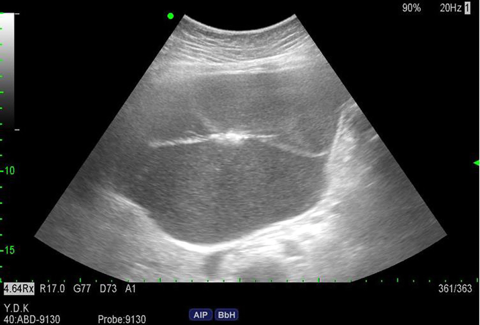
Fig. 1. Abdomen ultrasonography shows a large cystic mass with septae in liver left lobe.
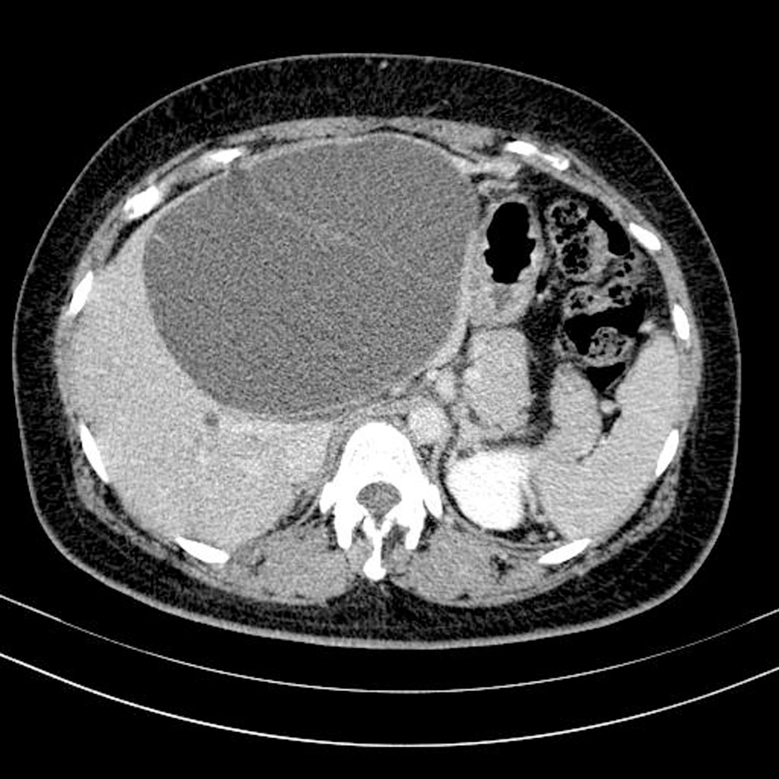
Fig. 2. Computed tomography shows a large cystic lesion with multiple-enhancing septae (arrow).
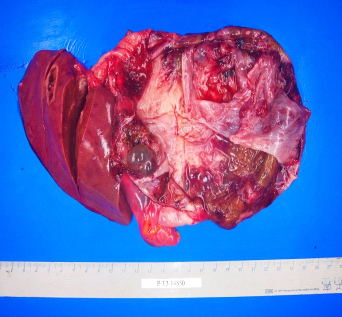
Fig. 3. Postoperative picture shows large thick walled cystic lesion in left lobe of liver.

Fig. 4. Cyst histology typical of a mucinous cystadenoma(A) Biliary cyst shows ovary like stroma (H &E stain, x400). (B) Arrow demonstrating ovary like stroma.
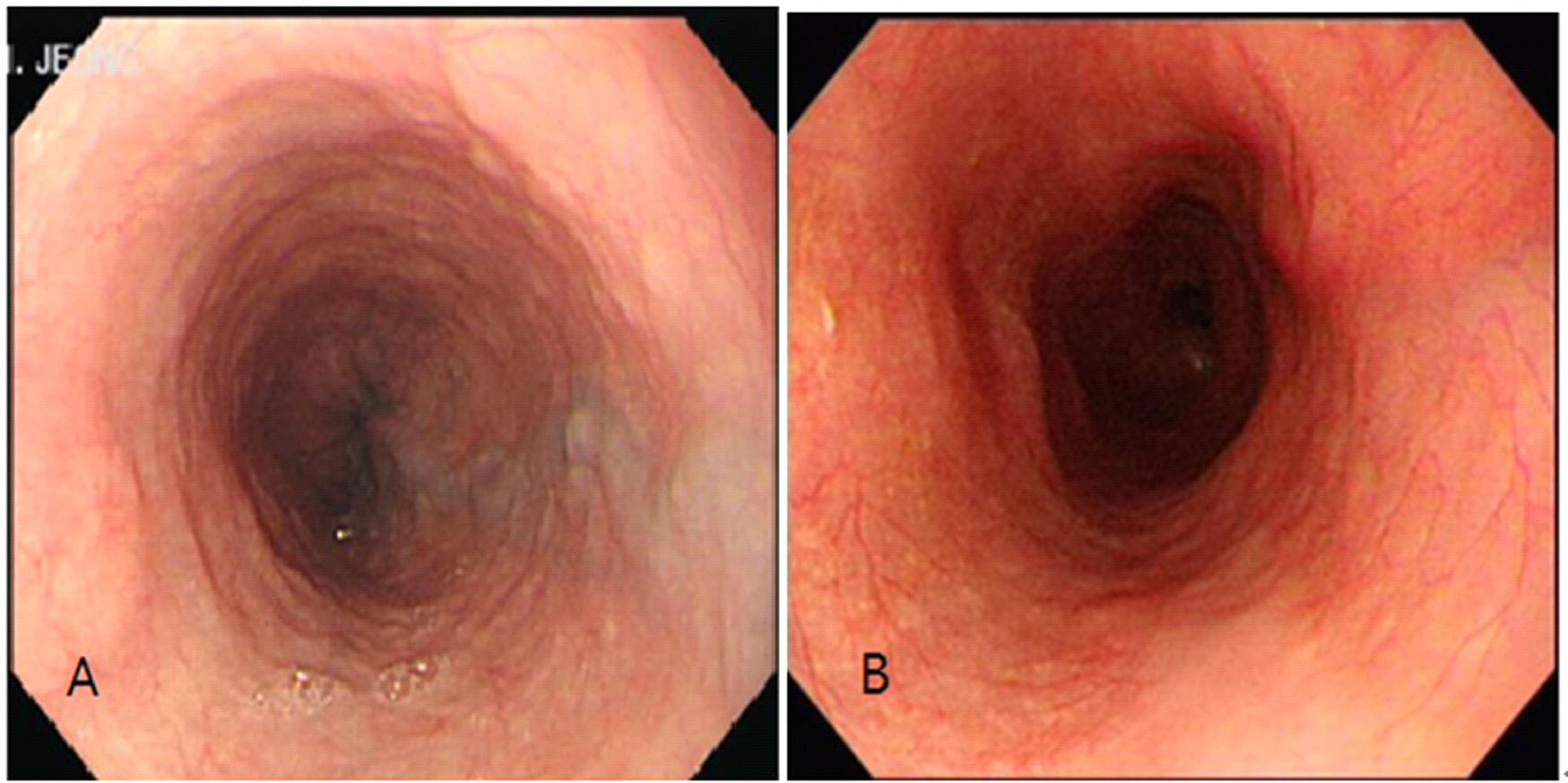
Fig. 5. Endoscopy image before operation demonstrating esophageal varix upto midesophagus.(A) Follow up endoscopy after lobectomy shows disappearance of esophageal varix.
Reference
-
References
1. Del Poggio P, Buonocore M. Cystic tumors of the liver: a practical approach. World J Gastroenterol. 2008; 14:3616–20.2. Ratti F, Ferla F, Paganelli M, Cipriani F, Aldrighetti L, Ferla G. Biliary cystadenoma: short- and longterm outcome after radical hepatic resection. Updates Surg. 2012; 64:13–8.
Article3. Grubor NM, Colovic RB, Atkinson HD, Micev MT. Giant biliary mucinous cystadenoma of the liver. Ann Hepatol. 2013; 12:979–83.
Article4. Erdogan D, Kloek J, Lamers WH, Offerhaus GJ, Busch OR, Gouma DJ, et al. Mucinous cystadenomas in liver: management and origin. Dig Surg. 2010; 27:19–23.
Article5. Gamblin TC, Holloway SE, Heckman JT, Geller DA. Laparoscopic resection of benign hepatic cysts: a new standard. J Am Coll Surg. 2008; 207:731–6.
Article6. Choi HK, Lee JK, Lee KH, Lee KT, Rhee JC, Kim KH, et al. Differential diagnosis for intrahepatic biliary cystadenoma and hepatic simple cyst: significance of cystic fluid analysis and radiologic findings. J Clin Gastroenterol. 2010; 44:289–93.7. Ryu KH, Lee TH, Kwon TG. Vascular lesion in an adult mimicking esophageal varix. Gastroenterololy. 2013; 144:35.
Article
- Full Text Links
-
- Actions
-
Cited
- CITED
-
- Close
- Share
-
- Similar articles
-
- Esophageal Motility and Acid Clearance in Patients with Esophageal Varices
- A Study of Endoscopic Variceal Ligation of under 6-Year-Old Aged Children with Esophageal Varices
- Effect of pro-banthine in diagnosis of esophageal varices
- Pseudotumoral gastric varices
- Duodenal Varices Causing Massive Upper Gastrointestinal Hemorrhage
