Hip Pelvis.
2016 Dec;28(4):264-268. 10.5371/hp.2016.28.4.264.
Femur Neck Fracture in a Young Marfan Syndrome Patient
- Affiliations
-
- 1Department of Orthopaedic Surgery, Busan Paik Hospital, Inje University College of Medicine, Busan, Korea. slowsting3@naver.com
- KMID: 2364706
- DOI: http://doi.org/10.5371/hp.2016.28.4.264
Abstract
- Marfan syndrome is an autosomal dominant and could decrease bone mineral density. So patients with Marfan syndrome could vulnerable to trauma in old ages. We present the first report, to the best of our knowledge, of a rare fracture of the femoral neck with a minor traumatic history in a juvenile Marfan syndrome patient whose physis is still open. Although the patient is young, her bone mineral density was low and the geometry of femur is changed like old ages. The femur neck fracture in children is very rare and only caused by high energy trauma, we concluded that the Marfan syndrome makes the bone weaker in young age and preventative medications to avoid fractures in younger Marfan syndrome patients are necessary in early ages.
Keyword
MeSH Terms
Figure
-
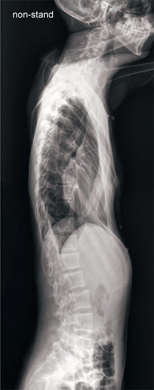
Fig. 1 Prominent pectus excavatum upon lateral chest X-ray.

Fig. 2 Pelvic anteroposterior X-ray demonstrating the transcervical fracture of the neck of the right femur.

Fig. 3 Initial chest X-ray showing scoliosis and a Cobb’s angle of 27° (T10-L4).
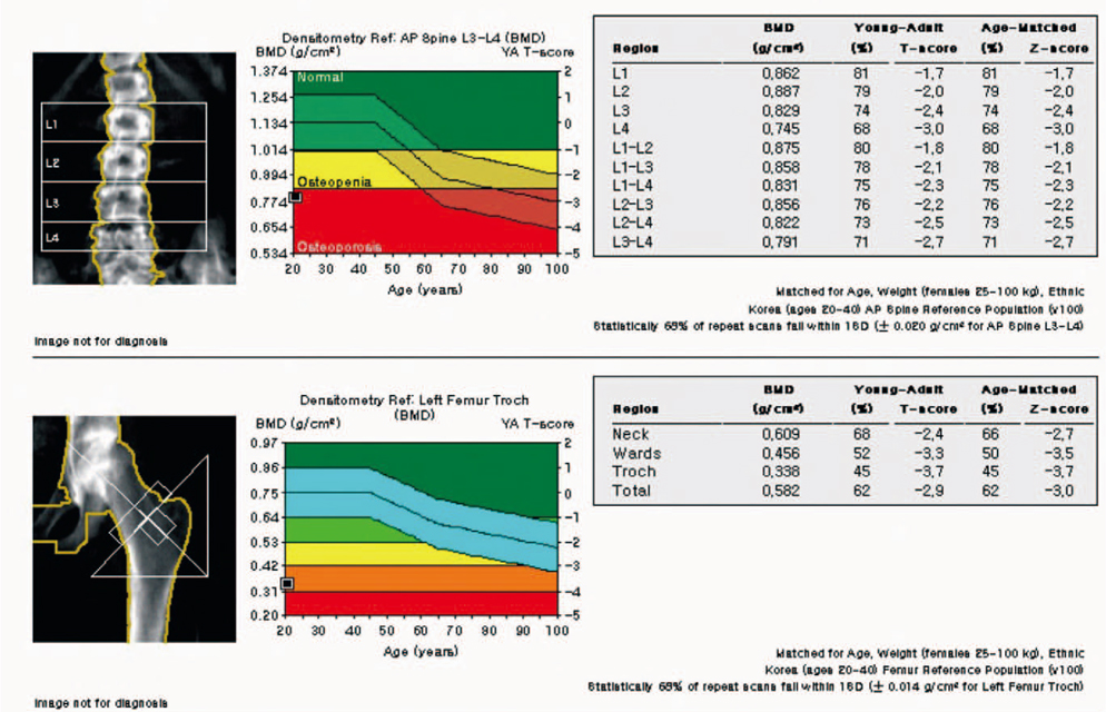
Fig. 4 Initial bone mineral density test showing osteoporosis in both the spine and femur.

Fig. 5 Post-operative X-ray showing stable fixation.
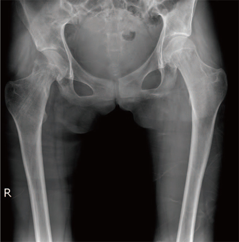
Fig. 6 X-ray showing good union after hardware removal, without displacement, malalignment, or avascular necrosis of the femoral head.
Cited by 1 articles
-
Atraumatic Bilateral Fracture of the Femoral Neck in Young Male Patient with Suspected Osteomalacia
Byung-Ho Yoon, Min-Soo Kwon
J Bone Metab. 2017;24(3):197-200. doi: 10.11005/jbm.2017.24.3.197.
Reference
-
1. Hindocha S, Kershaw S, Clayson AD. Atraumatic fracture neck of femur in Marfan’s syndrome: a case report. Injury Extra. 2007; 38:343–345.
Article2. Moura B, Tubach F, Sulpice M, et al. Bone mineral density in Marfan syndrome. A large case-control study. Joint Bone Spine. 2006; 73:733–735.
Article3. Alonso CG, Curiel MD, Carranza FH, Cano RP, Peréz AD. Femoral bone mineral density, neck-shaft angle and mean femoral neck width as predictors of hip fracture in men and women. Multicenter Project for Research in Osteoporosis. Osteoporos Int. 2000; 11:714–720.4. Avivi E, Arzi H, Paz L, Caspi I, Chechik A. Skeletal manifestations of marfan syndrome. IMAJ. 2008; 10:186–188.5. Boardman MJ, Herman MJ, Buck B, Pizzutillo PD. Hip fractures in children. J Am Acad Orthop Surg. 2009; 17:162–173.
Article6. Le Parc JM, Plantin P, Jondeau G, Goldschild M, Albert M, Boileau C. Bone mineral density in sixty adult patients with Marfan syndrome. Osteoporos Int. 1999; 10:475–479.
Article7. Kohlmeier L, Gasner C, Bachrach LK, Marcus R. The bone mineral status of patients with Marfan syndrome. J Bone Miner Res. 1995; 10:1550–1555.
Article8. Shin YL. Assessment of bone mineral density. J Korean Soc Pediatr Endocrinol. 2006; 11:123–130.9. Lee HJ, Song BS, Kim DH, et al. Bone mineral density reference of 10-20 year-old Korean children and adolescents: based on hologic DXA from the Korean national health and nutrition examination surveys. J Korean Soc Pediatr Endocrinol. 2011; 16:92–99.
Article10. Oh YJ, La KS, Rhie YJ, et al. Bone mineral density and correlation factors in normal children and adolescence. J Korean Soc Pediatr Endocrinol. 2009; 14:38–44.
- Full Text Links
-
- Actions
-
Cited
- CITED
-
- Close
- Share
-
- Similar articles
-
- Bilateral Femoral Neck Fractures in a Young Adult: A Case Report
- Fracture of Femur Neck with Heterotopic Ossification in Spinal Cord Injured Patient
- Femur neck fracture during open intramedullary nailing of femur shaft fracture: a report of one case
- Femur Neck Fracture during Closed Medullary Nailing of Femur Shaft Fracture: A Report of Two Cases
- Insufficiency Fracture of Ipsilateral Femur Neck in Patient Treated with Long Term Bisphosphonate Treatment: A Case Report
