Treatment of osteomyelitis in the rear area of the lingula of the mandible using sagittal split ramus osteotomy: a case report
- Affiliations
-
- 1Department of Oral and Maxillofacial Surgery, Busan Paik Hospital, Inje University College of Medicine, Busan, Korea. dwjty@hanmail.net
- KMID: 2364018
- DOI: http://doi.org/10.5125/jkaoms.2015.41.4.203
Abstract
- Osteomyelitis is classified into three groups according to its origin: osteomyelitis that originates from the blood supply, osteomyelitis related to bone disease or vascular disease, and osteomyelitis related to a local infection of dental or non-dental origin. The present case involved osteomyelitis related to a local infection of dental origin and was located in the rear area of the lingula of the mandible. We decided to use sagittal split ramus osteotomy to access the osteomyelitis area. Under general anesthesia, we successfully performed surgical sequestrectomy and curettage via sagittal split ramus osteotomy.
MeSH Terms
Figure
-
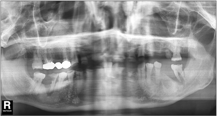
Fig. 1 Preoperative panorama X-ray. Severe alveolar bone loss around mandibular left second molar (#37) and multiple retained roots and dental caries of mandibular right first molar (#46) were observed.
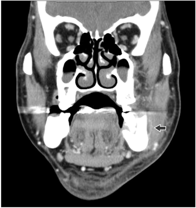
Fig. 2 Preoperative computed tomography. Pus formation (arrow) was observed left buccal and submassetric area.
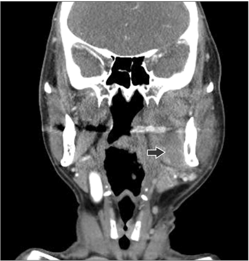
Fig. 3 Postoperative neck computed tomography. New appearance of immature abscess in left pterygomandibular and parapharyngeal space (2.5×5.1 cm; arrow).
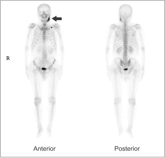
Fig. 4 Three phase bone scan. The enhancement (arrow) on the left mandible increased.
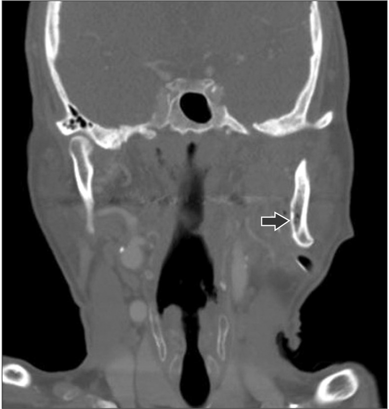
Fig. 5 Follow-up neck computed tomography. Findings of osteomyelitis (arrow) were observed on the rear area of lingula of the left mandible.
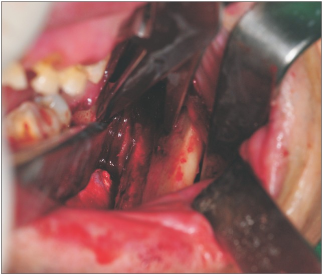
Fig. 6 Intraoperative intraoral photograph. We did surgical sequestrectomy and curettage successfully via the approach of sagittal split ramus osteotomy.
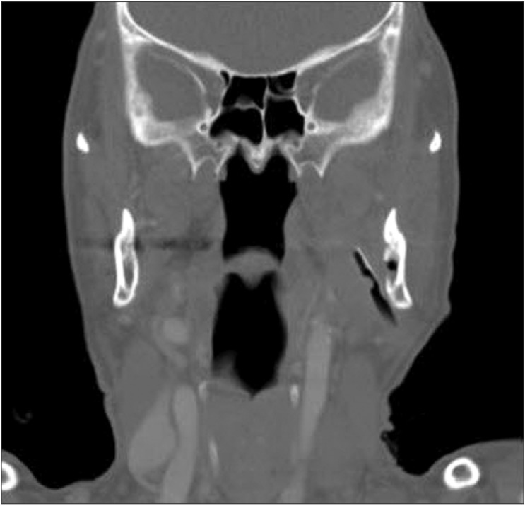
Fig. 7 Postoperative computed tomography. The operation was done successfully via the approach of sagittal split ramus osteotomy.
Reference
-
1. Korean Association of Oral and Maxillofacial Surgeons. Textbook of oral and maxillofacial surgery. 1st ed. Seoul: Dental and Medical Publishing Co.;1998.2. Kang HJ, Lee JH, Kim YD, Byun JH, Shin SH, Kim UK, et al. Osteomyelitis occuring in the zygoma caused by odontogenic maxillary sinusitis: case report. J Korean Assoc Oral Maxillofac Surg. 2004; 30:251–254.3. Kim SK, Sohn DS, Go MS, Seo JS, Lee CH. Treatment of osteomyelitis caused by fracture of the mandible. J Korean Assoc Maxillofac Plast Reconstr Surg. 1995; 17:277–282.4. Heo NO, Park JH, Shin YG, Pang SJ, Jeon IS, Yoon KH. A case of the treatment of osteomyelitis following open reduction of mandibular fracture. J Korean Assoc Maxillofac Plast Reconstr Surg. 1996; 18:712–717.5. Lee DJ, Choi MK, Oh SH, Lee JB. Conservative treatment of chronic suppurative osteomyelitis on mandibular body to condyle area: a case report. J Korean Assoc Oral Maxillofac Surg. 2009; 35:474–480.
- Full Text Links
-
- Actions
-
Cited
- CITED
-
- Close
- Share
-
- Similar articles
-
- A Study Of Mandibular Anatomy For Orthognathic Surgery In Koreans
- Surgical correction of maxillofacial deformity with fibrous-osseous lesion of mandible using the intraoral vertical ramus osteotomy
- A case report of hemifacial microsomia
- Mandibular anatomy related to sagittal split ramus osteotomy in Koreans
- Retromandibular vein position and course patterns in relation to mandible: anatomical morphologies requiring particular vigilance during sagittal split ramus osteotomy
