Retromandibular vein position and course patterns in relation to mandible: anatomical morphologies requiring particular vigilance during sagittal split ramus osteotomy
- Sugahara K
 1,2
1,2 - Matsunaga S2,3
- Yamamoto M3
- Noguchi T3
- Morita S3
- Koyachi M1
- Koyama Y1
- Koyama T1
- Kasahara N4
- Abe S2,3
- Katakura A1,2
- Affiliations
-
- 1Department of Oral Pathobiological Science and Surgery, Tokyo Dental College, Tokyo, Japan
- 2Oral Health Science Center, Tokyo Dental College, Tokyo, Japan
- 3Department of Anatomy, Tokyo Dental College, Tokyo, Japan
- 4Department of Forensic Odontology and Anthropology, Tokyo Dental College, Tokyo, Japan
- KMID: 2509690
- DOI: http://doi.org/10.5115/acb.20.236
Abstract
- Major bleeding associated with sagittal split ramus osteotomy (SSRO) involves vessels such as the inferior alveolar, facial, and maxillary arteries and veins, and the retromandibular vein (RMV). The present study aimed to clarify and classify the three-dimensional variations in RMV position and course direction in relation to the mandible. Specimens comprised a total of 15 scientific cadavers, and the relationship between RMV and the mandible lateral and posterior views was observed. We identified 3 patterns on the lateral view, the mean distance between the RMV and the posterior border of the ramus was 3.9 mm at the height of the lingula. A total of five course patterns were identified on the posterior view. In no course pattern, the RMV inferior to the lingula was lateral to its position superior to the lingual. The present findings suggest that it may be possible to predict correlations with intraoperative bleeding risk. Further study is planned using contrast computed tomography in patients with jaw deformity for skeletal classification.
Keyword
Figure
-
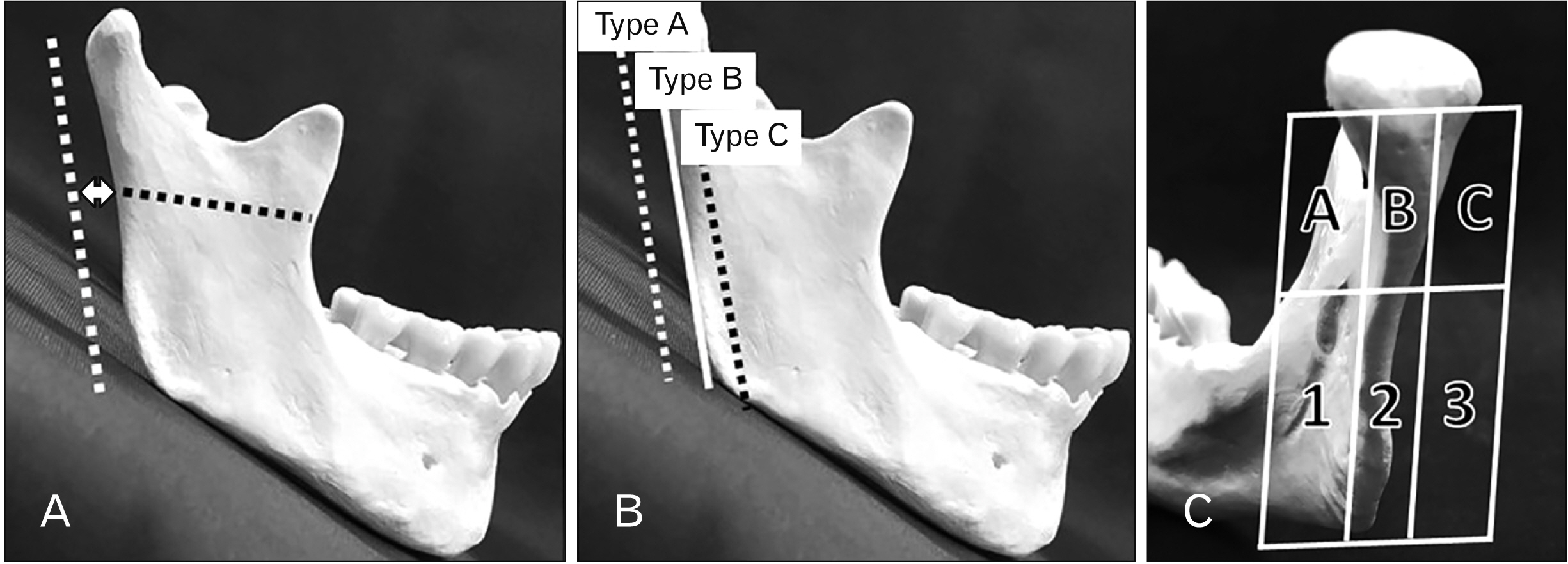
Fig. 1 (A) Measurement distance between the posterior border of the mandibular ramus and RMV at the height of the mandibular lingula. Dotted white line: RMV; dotted black line: height of the mandibular lingula; arrow: measurement distance. (B) Classifications of the positional relationship between RMV and the posterior border of the mandibular ramus from a lateral view. (Type A) RMV positioned posterior to the posterior border of the ramus, (Type B) RMV adjoining the posterior border of the ramus, (Type C) RMV positioned anterior to the posterior border of the ramus. (C) RMV course patterns on the posterior view of the mandibular ramus. Images were divided into three buccolingual sections in relation to the ramus. These were then each divided horizontally at the height of the mandibular lingula. The upper part is divided into three parts (A, B, C), and the lower part is divided into three parts (1, 2, 3). The driving was classified running pattern by combining one each in the upper and lower parts. RMV, retromandibular vein.
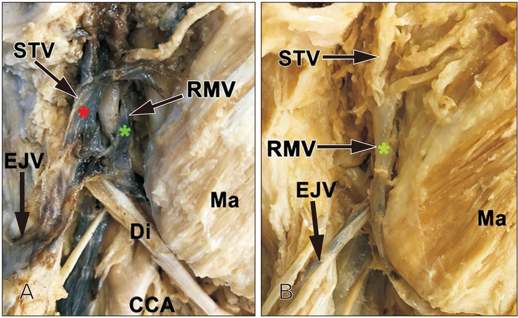
Fig. 2 Topographical anatomy of the retromandibular region. (Panel A) The RMV (green star) and the STV (red star) run posteriorly to the mandibular ramus (86.7%). (Panel B) The superior (inferior) part of the RMV (green star) is continuous with the STV (the internal jugular vein) (13.3%). CCA, common carotid artery; Di, digastric muscle; EJV, external jugular vein; M, mandibular bone; Ma, masseter; RMV, retromandibular vein; STV, superficial temporal vein.
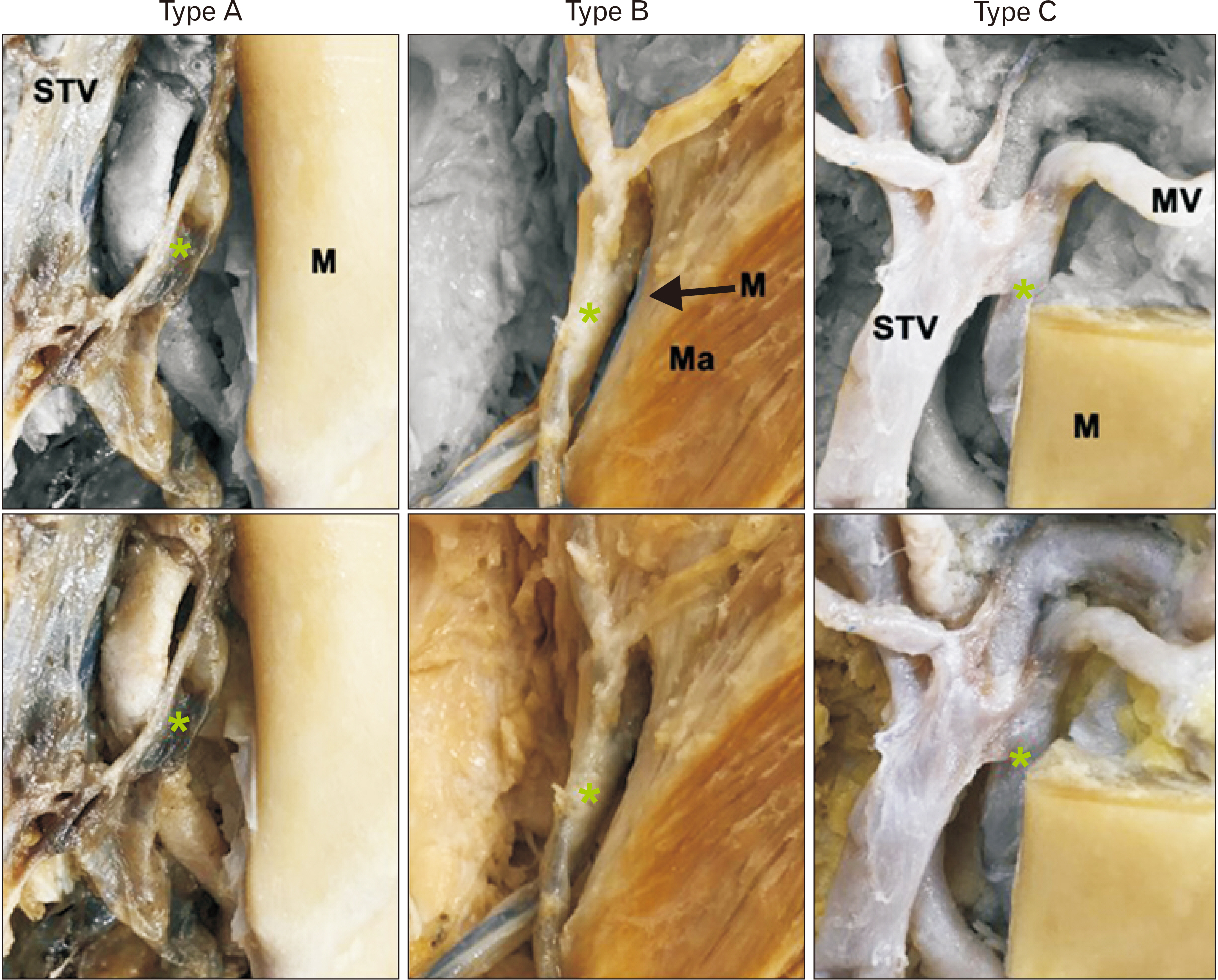
Fig. 3 Positioning of the posterior border of the mandible and posterior veins of the mandible. Panel A–C inverts the vein of Panel A’–C’ (original image) and the other than the mandible (or cusp). (Type A) The posterior border of the mandible is separated from the posterior vein of the mandible (Panel A and A’). (Type B) The anterior border of the mandibular posterior vein is in contact with the posterior border of the mandibular branch (Panel B and B’). (Type C) The posterior vein of the mandible is located in front of the posterior border of the mandible and inside the posterior border of the mandible (Panel C and C’). Asterisk, retromandibular vein; M, mandibular bone; Ma, masseter; STV, superficial temporal vein.

Fig. 4 Course pattern on the posterior view of RMV and the posterior border of the mandibular ramus. RMV, retromandibular vein.
Reference
-
References
1. Kobayashi T, Saito C, Inoue N, Ohata N, Kawamura H, Goto S, Goto M, Shiratsuchi Y, Susami T, Tanne K, Hashimoto K, Moriyama K, Amagasa T, Himuro T, Tonoki M. 2008; Treatment of jaw deformity: a nationwide survey of the situation in Japan. JPn J Jaw Deform. 18:237–50. DOI: 10.5927/jjjd1991.18.237.
Article2. Hamada Y, Sugahara K, Yoshida S, Watanabe A, Bessho H, Kasahara K, Takano M, Saito C, Shibahara T, Katakura A. 2019; A 27-year retrospective clinical analysis of 2640 orthognathic surgery cases in the Tokyo Dental College. J Oral Maxillofac Surg Med Pathol. 31:305–10. DOI: 10.1016/j.ajoms.2019.03.008.
Article3. O'Ryan F. 1990; Complications of orthognathic surgery. Oral Maxillofac Surg Clin North Am. 2:593–613. PMID: 2680224.4. Sahoo NK, Kaur P, Sharma IDRR. 2017; Complications of sagittal split ramus osteotomy. J Oral Maxillofac Surg Med Pathol. 29:100–4. DOI: 10.1016/j.ajoms.2016.09.006.
Article5. Odaka K, Matsunaga S. 2020; Course of the maxillary vein and its positional relationship with the mandibular ramus require attention during mandibuloplasty. J Craniofac Surg. 31:861–4. DOI: 10.1097/SCS.0000000000006174. PMID: 31842072. PMCID: PMC7329198.
Article6. Norton NS. 2016. Netter's head and neck anatomy for dentistry. 3rd ed. Elsevier;Philadelphia:7. Touré G, Vacher C. 2010; Relations of the facial nerve with the retromandibular vein: anatomic study of 132 parotid glands. Surg Radiol Anat. 32:957–61. DOI: 10.1007/s00276-010-0674-9. PMID: 20473672.
Article8. Babademez MA, Acar B, Gunbey E, Karabulut H, Karasen RM. 2010; Anomalous relationship of the retromandibular vein to the facial nerve as a potential risk factor for facial nerve injury during parotidectomy. J Craniofac Surg. 21:801–2. DOI: 10.1097/SCS.0b013e3181d84027. PMID: 20485053.
Article9. Loukota RA, Eckelt U, De Bont L, Rasse M. 2005; Subclassification of fractures of the condylar process of the mandible. Br J Oral Maxillofac Surg. 43:72–3. DOI: 10.1016/j.bjoms.2004.08.018. PMID: 15620780.
Article10. Trauner R, Obwegeser H. 1957; The surgical correction of mandibular prognathism and retrognathia with consideration of genioplasty. I. Surgical procedures to correct mandibular prognathism and reshaping of the chin. Oral Surg Oral Med Oral Pathol. 10:677–89. contd. DOI: 10.1016/S0030-4220(57)80063-2. PMID: 13441284.11. Dal Pont G. 1961; Retromolar osteotomy for the correction of prognathism. J Oral Surg Anesth Hosp Dent Serv. 19:42–7. PMID: 13719390.12. Hunsuck EE. 1968; A modified intraoral sagittal splitting technic for correction of mandibular prognathism. J Oral Surg. 26:250–3. PMID: 5237786.13. Epker BN. 1977; Modifications in the sagittal osteotomy of the mandible. J Oral Surg. 35:157–9. PMID: 264514.14. Panula K, Finne K, Oikarinen K. 2001; Incidence of complications and problems related to orthognathic surgery: a review of 655 patients. J Oral Maxillofac Surg. 59:1128–36. discussion 1137DOI: 10.1053/joms.2001.26704. PMID: 11573165.
Article15. Patel PK, Morris DE, Gassman A. 2007; Complications of orthognathic surgery. J Craniofac Surg. 18:975–85. Quiz 986–8. DOI: 10.1097/scs.0b013e318068442c. PMID: 17667699.
Article16. Lanigan DT, Hey JH, West RA. 1991; Major vascular complications of orthognathic surgery: false aneurysms and arteriovenous fistulas following orthognathic surgery. J Oral Maxillofac Surg. 49:571–7. DOI: 10.1016/0278-2391(91)90337-L. PMID: 2037912.
Article17. Piñeiro-Aguilar A, Somoza-Martín M, Gandara-Rey JM, García-García A. 2011; Blood loss in orthognathic surgery: a systematic review. J Oral Maxillofac Surg. 69:885–92. DOI: 10.1016/j.joms.2010.07.019. PMID: 21195531.
Article18. Kim SG, Park SS. 2007; Incidence of complications and problems related to orthognathic surgery. J Oral Maxillofac Surg. 65:2438–44. DOI: 10.1016/j.joms.2007.05.030. PMID: 18022466.
Article19. Mei A, Qiu L. 2019; The efficacy of tranexamic acid for orthognathic surgery: a meta-analysis of randomized controlled trials. Int J Oral Maxillofac Surg. 48:1323–8. DOI: 10.1016/j.ijom.2018.07.027. PMID: 30902548.
Article20. Olsen JJ, Skov J, Ingerslev J, Thorn JJ, Pinholt EM. 2016; Prevention of bleeding in orthognathic surgery--a systematic review and meta-analysis of randomized controlled trials. J Oral Maxillofac Surg. 74:139–50. DOI: 10.1016/j.joms.2015.05.031. PMID: 26073131.
Article21. Jackson IT, Jack CR, Aycock B, Dubin B, Irons GB. 1989; The management of intraosseous arteriovenous malformations in the head and neck area. Plast Reconstr Surg. 84:47–54. DOI: 10.1097/00006534-198907000-00010. PMID: 2734403.
Article22. Kallela I, Söderholm AL, Paukku P, Lindqvist C. 1995; Lag-screw osteosynthesis of mandibular condyle fractures: a clinical and radiological study. J Oral Maxillofac Surg. 53:1397–404. discussion 1405–6. DOI: 10.1016/0278-2391(95)90663-0. PMID: 7490649.
Article23. Handschel J, Rüggeberg T, Depprich R, Schwarz F, Meyer U, Kübler NR, Naujoks C. 2012; Comparison of various approaches for the treatment of fractures of the mandibular condylar process. J Craniomaxillofac Surg. 40:e397–401. DOI: 10.1016/j.jcms.2012.02.012. PMID: 22440318.
Article24. Devlin MF, Hislop WS, Carton AT. 2002; Open reduction and internal fixation of fractured mandibular condyles by a retromandibular approach: surgical morbidity and informed consent. Br J Oral Maxillofac Surg. 40:23–5. DOI: 10.1054/bjom.2001.0748. PMID: 11883965.25. Ellis E 3rd, McFadden D, Simon P, Throckmorton G. 2000; Surgical complications with open treatment of mandibular condylar process fractures. J Oral Maxillofac Surg. 58:950–8. DOI: 10.1053/joms.2000.8734. PMID: 10981974.
Article
- Full Text Links
-
- Actions
-
Cited
- CITED
-
- Close
- Share
-
- Similar articles
-
- Treatment of osteomyelitis in the rear area of the lingula of the mandible using sagittal split ramus osteotomy: a case report
- Skeletal relapse pattern after sagittal split ramus osteotomy of mandibular prognathic patient
- A case report of hemifacial microsomia
- Enucleation of large keratocystic odontogenic tumor at mandible via unilateral sagittal split osteotomy: a report of three cases
- The postoperative condylar posit10n related to temporomandibular discomfort in sagittal split ramus osteotomy
