Clin Orthop Surg.
2016 Mar;8(1):92-98. 10.4055/cios.2016.8.1.92.
The Efficacy of Percutaneous Lateral Hemiepiphysiodesis on Angular Correction in Idiopathic Adolescent Genu Varum
- Affiliations
-
- 1Department of Orthopaedic Surgery, Samsung Medical Center, Sungkyunkwan University School of Medicine, Seoul, Korea. jss3505@skku.edu
- KMID: 2363951
- DOI: http://doi.org/10.4055/cios.2016.8.1.92
Abstract
- BACKGROUND
Percutaneous lateral hemiepiphysiodesis of the lower extremity is a simple and excellent method to correct the angular and length problems cosmetically. However, the efficacy of percutaneous lateral hemiepiphysiodesis is not well established in the literature. The purpose of this study was to evaluate the efficacy of percutaneous lateral hemiepiphysiodesis for angular corrections in adolescent idiopathic genu varum patients with proximal tibia vara and identify the factors affecting the amount of deformity correction of the lower limb in the coronal plane.
METHODS
We retrospectively reviewed 20 patients (40 lower limbs) who had percutaneous lateral hemiepiphysiodesis on the proximal lateral tibia between 1997 and 2010. Radiographic evaluations were made using (1) the hip-knee-ankle angle and (2) the length of the tibia. Furthermore, the intercondylar distance was evaluated at the level of the knee joint. Preoperative factors (gender, age, body mass index, intercondylar distance, preoperative hip-knee-ankle angle, remaining growth of tibia, and calculated correctable angle) were analyzed, as well as their correlation with the degree of the actual correction angle.
RESULTS
The amount of coronal deformity of the lower limb was improved from its preoperative state. The median average of hip-knee-ankle angle improved from 8.0degrees (interquartile range [IQR], 7.0degrees to 10.0degrees) preoperatively to 3.0degrees (IQR, 2.5degrees to 4.0degrees) at the final follow-up (p < 0.001). The median percent ratio of the angular correction was 60% (IQR, 50% to 71.3%). The correlation coefficients were -0.537, 0.832, 0.791, and 0.685 for the bone age, preoperative hip-knee-ankle angle, the remaining growth of tibia, and calculated correctable angle, respectively.
CONCLUSIONS
Despite the excellent cosmetic outcome of percutaneous lateral hemiepiphysiodesis on the proximal lateral tibia in adolescent idiopathic genu varum, the effect was limited in most cases. For optimum results, surgery a few months earlier is recommended, rather than at the calculated operation time.
MeSH Terms
Figure
-
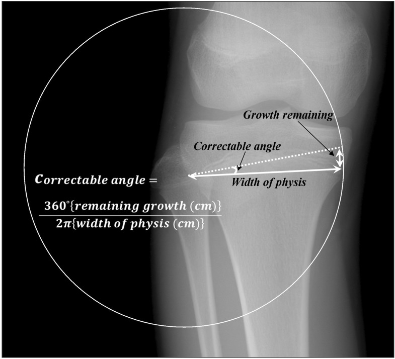
Fig. 1 Schematic presentation for the correctable angle by asymmetrical lateral proximal tibia epiphysiodesis.
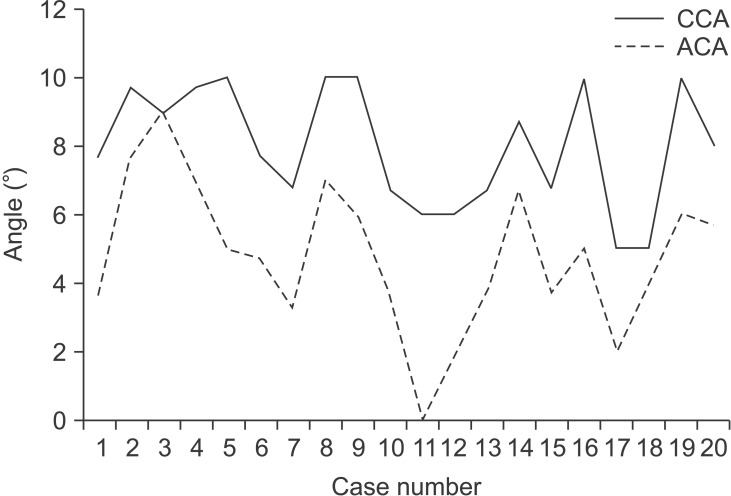
Fig. 2 Comparison of the correction angle between the calculated expectation values and actual values. CCA: calculated correctable angle, ACA: actual correction angle at final follow-up.
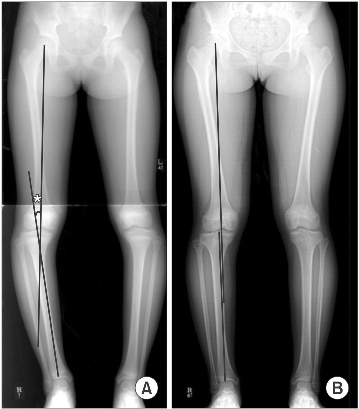
Fig. 3 Case number 3. A 10.3-year-old girl (bone age). (A) Preoperative radiograph. (B) Perfect alignment was achieved 2 years postoperatively when skeletal maturity was reached. *Hip-knee-ankle angle.
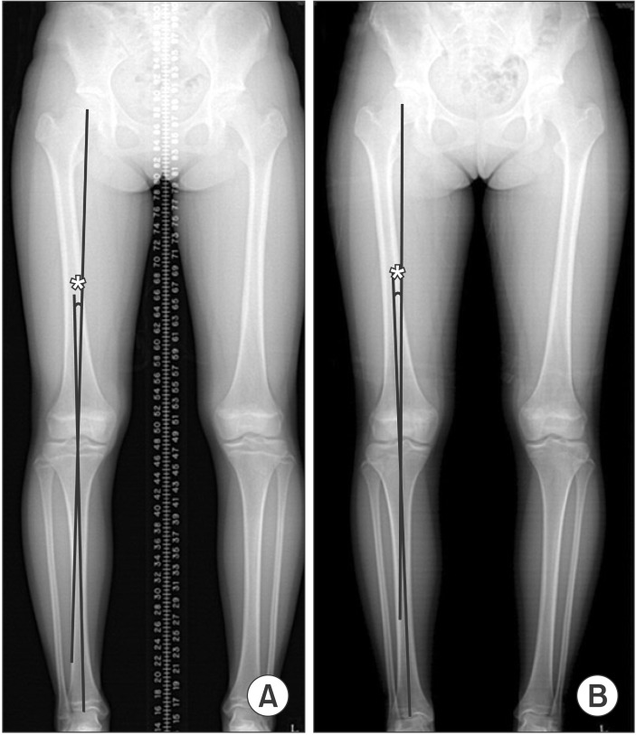
Fig. 4 Case number 11. A 12.3-year-old girl (bone age). (A) Preoperative radiograph. (B) There was no improvement of angular deformity at final follow-up. *Hip-knee-ankle angle.
Reference
-
1. Bowen JR, Leahey JL, Zhang ZH, MacEwen GD. Partial epiphysiodesis at the knee to correct angular deformity. Clin Orthop Relat Res. 1985; (198):184–190. PMID: 4028549.
Article2. Babu LV, Evans O, Sankar A, Davies AG, Jones S, Fernandes JA. Epiphysiodesis for limb length discrepancy: a comparison of two methods. Strategies Trauma Limb Reconstr. 2014; 9(1):1–3. PMID: 24271553.3. Horn J, Gunderson RB, Wensaas A, Steen H. Percutaneous epiphysiodesis in the proximal tibia by a single-portal approach: evaluation by radiostereometric analysis. J Child Orthop. 2013; 7(4):295–300. PMID: 24432090.
Article4. Ferrick MR, Birch JG, Albright M. Correction of non-Blount's angular knee deformity by permanent hemiepiphyseodesis. J Pediatr Orthop. 2004; 24(4):397–402. PMID: 15205622.
Article5. Anderson M, Green WT, Messner MB. Growth and predictions of growth in the lower extremities. J Bone Joint Surg Am. 1963; 45-A(1):1–14. PMID: 14040773.
Article6. Lee SC, Shim JS, Seo SW, Lim KS, Ko KR. The accuracy of current methods in determining the timing of epiphysiodesis. Bone Joint J. 2013; 95-B(7):993–1000. PMID: 23814256.
Article7. Sheehy L, Felson D, Zhang Y, et al. Does measurement of the anatomic axis consistently predict hip-knee-ankle angle (HKA) for knee alignment studies in osteoarthritis? Analysis of long limb radiographs from the multicenter osteoarthritis (MOST) study. Osteoarthritis Cartilage. 2011; 19(1):58–64. PMID: 20950695.
Article8. Yoshioka Y, Siu DW, Scudamore RA, Cooke TD. Tibial anatomy and functional axes. J Orthop Res. 1989; 7(1):132–137. PMID: 2908904.
Article9. Inan M, Chan G, Bowen JR. Correction of angular deformities of the knee by percutaneous hemiepiphysiodesis. Clin Orthop Relat Res. 2007; 456:164–169. PMID: 17065845.
Article10. Schroerlucke S, Bertrand S, Clapp J, Bundy J, Gregg FO. Failure of Orthofix eight-Plate for the treatment of Blount disease. J Pediatr Orthop. 2009; 29(1):57–60. PMID: 19098648.
Article11. Stevens PM. Guided growth for angular correction: a preliminary series using a tension band plate. J Pediatr Orthop. 2007; 27(3):253–259. PMID: 17414005.12. Shin SJ, Cho TJ, Park MS, et al. Angular deformity correction by asymmetrical physeal suppression in growing children: stapling versus percutaneous transphyseal screw. J Pediatr Orthop. 2010; 30(6):588–593. PMID: 20733425.13. Wiemann JM 4th, Tryon C, Szalay EA. Physeal stapling versus 8-plate hemiepiphysiodesis for guided correction of angular deformity about the knee. J Pediatr Orthop. 2009; 29(5):481–485. PMID: 19568021.
Article14. Khoury JG, Tavares JO, McConnell S, Zeiders G, Sanders JO. Results of screw epiphysiodesis for the treatment of limb length discrepancy and angular deformity. J Pediatr Orthop. 2007; 27(6):623–628. PMID: 17717460.
Article15. Blount WP. A mature look at epiphyseal stapling. Clin Orthop Relat Res. 1971; 77:158–163. PMID: 5140445.16. Metaizeau JP, Wong-Chung J, Bertrand H, Pasquier P. Percutaneous epiphysiodesis using transphyseal screws (PETS). J Pediatr Orthop. 1998; 18(3):363–369. PMID: 9600565.
- Full Text Links
-
- Actions
-
Cited
- CITED
-
- Close
- Share
-
- Similar articles
-
- Contributions of the Distal Femur and Proximal Tibia to Idiopathic Genu Varum and Genu Valgum in Adolescents
- Genu Varum, Both
- Change in Effective Leg Length after Angular Deformity Correction by Hemiepiphyseal Stapling
- Deformity Correction of the Knee and Leg Lengthening by the Ilizarov Method in Children with Vitamin D Resistant Rickets
- Pelvic, Hip, and Knee Kinematics of Stair Climbing in People with Genu Varum
