J Korean Med Sci.
2016 Mar;31(3):389-396. 10.3346/jkms.2016.31.3.389.
Elevated Erythrocyte Sedimentation Rate Is Predictive of Interstitial Lung Disease and Mortality in Dermatomyositis: a Korean Retrospective Cohort Study
- Affiliations
-
- 1Department of Internal Medicine, Seoul National University Hospital, Seoul, Korea. jinkyunpark@gmail.com
- 2Department of Molecular Medicine and Biopharmaceutical Sciences, Graduate School of Convergence Science and Technology, and College of Medicine, Medical Research Institute, Seoul National University, Seoul, Korea.
- 3Department of Medicine, Johns Hopkins University School of Medicine, Baltimore, MD, USA.
- KMID: 2363499
- DOI: http://doi.org/10.3346/jkms.2016.31.3.389
Abstract
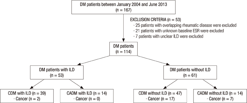
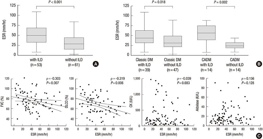
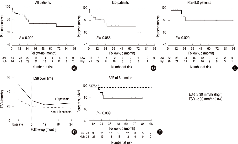
- Interstitial lung disease (ILD) is a major cause of death in patients with dermatomyositis (DM). This study was aimed to examine the utility of the erythrocyte sedimentation rate (ESR) as a predictor of ILD and prognostic marker of mortality in patients with DM. One hundred-and-fourteen patients with DM were examined, including 28 with clinically amyopathic DM (CADM). A diagnosis of ILD was made based on high resolution computed tomography (HRCT) scans. The association between elevated ESR and pulmonary impairment and mortality was then examined. ILD was diagnosed in 53 (46.5%) of 114 DM patients. Cancer was diagnosed in 2 (3.8%) of 53 DM patients with ILD and in 24 (92.3%) of those without ILD (P < 0.001). The median ESR (50.0 mm/hour) in patients with ILD was significantly higher than that in patients without ILD (29.0 mm/hour; P < 0.001). ESR was inversely correlated with forced vital capacity (Spearman rho = - 0.303; P = 0.007) and carbon monoxide diffusing capacity (rho = - 0.319; P = 0.006). DM patients with baseline ESR > or = 30 mm/hour had significantly higher mortality than those with ESR < 30 mm/hour (P = 0.002, log-rank test). Patients with a persistently high ESR despite immunosuppressive therapy was associated with higher mortality than those with a normalized ESR (P = 0.039, log-rank test). Elevated ESR is associated with increased mortality in patients with DM due to respiratory failure. Thus, monitoring ESR should be an integral part of the clinical care of DM patients.
MeSH Terms
-
Adult
Asian Continental Ancestry Group
Blood Sedimentation
Carbon Monoxide/metabolism
Cohort Studies
Dermatomyositis/blood/*diagnosis/mortality
Disease Progression
Erythrocytes/*cytology
Female
Follow-Up Studies
Humans
Immunosuppressive Agents/therapeutic use
Lung Diseases, Interstitial/*complications/diagnosis
Male
Middle Aged
Predictive Value of Tests
Prognosis
Republic of Korea
Respiratory Function Tests
Retrospective Studies
Risk Factors
Survival Analysis
Carbon Monoxide
Immunosuppressive Agents
Figure
Reference
-
1. Marie I, Hatron PY, Hachulla E, Wallaert B, Michon-Pasturel U, Devulder B. Pulmonary involvement in polymyositis and in dermatomyositis. J Rheumatol. 1998; 25:1336–1343.2. Marie I, Hachulla E, Chérin P, Dominique S, Hatron PY, Hellot MF, Devulder B, Herson S, Levesque H, Courtois H. Interstitial lung disease in polymyositis and dermatomyositis. Arthritis Rheum. 2002; 47:614–622.3. Fathi M, Dastmalchi M, Rasmussen E, Lundberg IE, Tornling G. Interstitial lung disease, a common manifestation of newly diagnosed polymyositis and dermatomyositis. Ann Rheum Dis. 2004; 63:297–301.4. Sontheimer RD. Would a new name hasten the acceptance of amyopathic dermatomyositis (dermatomyositis siné myositis) as a distinctive subset within the idiopathic inflammatory dermatomyopathies spectrum of clinical illness? J Am Acad Dermatol. 2002; 46:626–636.5. Gerami P, Schope JM, McDonald L, Walling HW, Sontheimer RD. A systematic review of adult-onset clinically amyopathic dermatomyositis (dermatomyositis siné myositis): a missing link within the spectrum of the idiopathic inflammatory myopathies. J Am Acad Dermatol. 2006; 54:597–613.6. Mukae H, Ishimoto H, Sakamoto N, Hara S, Kakugawa T, Nakayama S, Ishimatsu Y, Kawakami A, Eguchi K, Kohno S. Clinical differences between interstitial lung disease associated with clinically amyopathic dermatomyositis and classic dermatomyositis. Chest. 2009; 136:1341–1347.7. Kang EH, Lee EB, Shin KC, Im CH, Chung DH, Han SK, Song YW. Interstitial lung disease in patients with polymyositis, dermatomyositis and amyopathic dermatomyositis. Rheumatology (Oxford). 2005; 44:1282–1286.8. Nawata Y, Kurasawa K, Takabayashi K, Miike S, Watanabe N, Hiraguri M, Kita Y, Kawai M, Saito Y, Iwamoto I. Corticosteroid resistant interstitial pneumonitis in dermatomyositis/polymyositis: prediction and treatment with cyclosporine. J Rheumatol. 1999; 26:1527–1533.9. Kobayashi N, Takezaki S, Kobayashi I, Iwata N, Mori M, Nagai K, Nakano N, Miyoshi M, Kinjo N, Murata T, et al. Clinical and laboratory features of fatal rapidly progressive interstitial lung disease associated with juvenile dermatomyositis. Rheumatology (Oxford). 2015; 54:784–791.10. Gono T, Kaneko H, Kawaguchi Y, Hanaoka M, Kataoka S, Kuwana M, Takagi K, Ichida H, Katsumata Y, Ota Y, et al. Cytokine profiles in polymyositis and dermatomyositis complicated by rapidly progressive or chronic interstitial lung disease. Rheumatology (Oxford). 2014; 53:2196–2203.11. Brigden ML. Clinical utility of the erythrocyte sedimentation rate. Am Fam Physician. 1999; 60:1443–1450.12. Amerio P, Girardelli CR, Proietto G, Forleo P, Cerritelli L, Feliciani C, Colonna L, Teofoli P, Amerio P, Puddu P, et al. Usefulness of erythrocyte sedimentation rate as tumor marker in cancer associated dermatomyositis. Eur J Dermatol. 2002; 12:165–169.13. Mallya RK, de Beer FC, Berry H, Hamilton ED, Mace BE, Pepys MB. Correlation of clinical parameters of disease activity in rheumatoid arthritis with serum concentration of C-reactive protein and erythrocyte sedimentation rate. J Rheumatol. 1982; 9:224–228.14. Park JK, Gelber AC, George M, Danoff SK, Qubti MA, Christopher-Stine L. Pulmonary impairment, not muscle injury, is associated with elevated ESR in the idiopathic inflammatory myopathies. Rheumatology (Oxford). 2013; 52:1336–1338.15. Bohan A, Peter JB, Bowman RL, Pearson CM. Computer-assisted analysis of 153 patients with polymyositis and dermatomyositis. Medicine (Baltimore). 1977; 56:255–286.16. Borawski J, Myśliwiec M. The hematocrit-corrected erythrocyte sedimentation rate can be useful in diagnosing inflammation in hemodialysis patients. Nephron. 2001; 89:381–383.17. Ikezoe J, Johkoh T, Kohno N, Takeuchi N, Ichikado K, Nakamura H. High-resolution CT findings of lung disease in patients with polymyositis and dermatomyositis. J Thorac Imaging. 1996; 11:250–259.18. Mino M, Noma S, Taguchi Y, Tomii K, Kohri Y, Oida K. Pulmonary involvement in polymyositis and dermatomyositis: sequential evaluation with CT. AJR Am J Roentgenol. 1997; 169:83–87.19. Moshage H. Cytokines and the hepatic acute phase response. J Pathol. 1997; 181:257–266.20. Sun Y, Liu Y, Yan B, Shi G. Interstitial lung disease in clinically amyopathic dermatomyositis (CADM) patients: a retrospective study of 41 Chinese Han patients. Rheumatol Int. 2013; 33:1295–1302.21. Park JK, Yoo HG, Ahn DS, Jeon HS, Yoo WH. Successful treatment for conventional treatment-resistant dermatomyositis-associated interstitial lung disease with adalimumab. Rheumatol Int. 2012; 32:3587–3590.22. Becker GJ, Waldburger M, Hughes GR, Pepys MB. Value of serum C-reactive protein measurement in the investigation of fever in systemic lupus erythematosus. Ann Rheum Dis. 1980; 39:50–52.23. Kirou KA, Lee C, George S, Louca K, Peterson MG, Crow MK. Activation of the interferon-alpha pathway identifies a subgroup of systemic lupus erythematosus patients with distinct serologic features and active disease. Arthritis Rheum. 2005; 52:1491–1503.24. Baechler EC, Bauer JW, Slattery CA, Ortmann WA, Espe KJ, Novitzke J, Ytterberg SR, Gregersen PK, Behrens TW, Reed AM. An interferon signature in the peripheral blood of dermatomyositis patients is associated with disease activity. Mol Med. 2007; 13:59–68.25. Walsh RJ, Kong SW, Yao Y, Jallal B, Kiener PA, Pinkus JL, Beggs AH, Amato AA, Greenberg SA. Type I interferon-inducible gene expression in blood is present and reflects disease activity in dermatomyositis and polymyositis. Arthritis Rheum. 2007; 56:3784–3792.26. Eloranta ML, Barbasso Helmers S, Ulfgren AK, Rönnblom L, Alm GV, Lundberg IE. A possible mechanism for endogenous activation of the type I interferon system in myositis patients with anti-Jo-1 or anti-Ro 52/anti-Ro 60 autoantibodies. Arthritis Rheum. 2007; 56:3112–3124.27. Sun WC, Sun YC, Lin H, Yan B, Shi GX. Dysregulation of the type I interferon system in adult-onset clinically amyopathic dermatomyositis has a potential contribution to the development of interstitial lung disease. Br J Dermatol. 2012; 167:1236–1244.28. Sato S, Hoshino K, Satoh T, Fujita T, Kawakami Y, Fujita T, Kuwana M. RNA helicase encoded by melanoma differentiation-associated gene 5 is a major autoantigen in patients with clinically amyopathic dermatomyositis: Association with rapidly progressive interstitial lung disease. Arthritis Rheum. 2009; 60:2193–2200.29. Marie I, Hachulla E, Hatron PY, Hellot MF, Levesque H, Devulder B, Courtois H. Polymyositis and dermatomyositis: short term and longterm outcome, and predictive factors of prognosis. J Rheumatol. 2001; 28:2230–2237.30. Chen YJ, Wu CY, Shen JL. Predicting factors of malignancy in dermatomyositis and polymyositis: a case-control study. Br J Dermatol. 2001; 144:825–831.31. Na SJ, Kim SM, Sunwoo IN, Choi YC. Clinical characteristics and outcomes of juvenile and adult dermatomyositis. J Korean Med Sci. 2009; 24:715–721.32. So MW, Koo BS, Kim YG, Lee CK, Yoo B. Idiopathic inflammatory myopathy associated with malignancy: a retrospective cohort of 151 Korean patients with dermatomyositis and polymyositis. J Rheumatol. 2011; 38:2432–2435.
- Full Text Links
- Actions
-
Cited
- CITED
-
- Close
- Share
- Similar articles
-
- A Case Of Dermatomyositis Associated With Combined Small Cell Lung Cancer
- Amyopathic Dermatomyositis with Interstitial Lung Disease: A Case Report
- A Case of Dermatomyositis with Acute Interstitial Pneumonitis Manifested as Antisynthetase Syndrome
- Acute Exacerbation of Interstitial Lung Disease in Newly Diagnosed Probable Dermatomyositis
- Rapidly Progressive Interstitial Lung Disease Associated with Dermatomyositis; A Case Report


