New method of assessing the relationship between buccal bone thickness and gingival thickness
- Affiliations
-
- 1Department of Periodontology, Seoul National University Gwanak Dental Hospital, Seoul, Korea.
- 2Department of Prosthodontics, Seoul National University Gwanak Dental Hospital, Seoul, Korea.
- 3Department of Periodontology, Dental Research Institute, Seoul National University School of Dentistry, Seoul, Korea. guy@snu.ac.kr
- KMID: 2362903
- DOI: http://doi.org/10.5051/jpis.2016.46.6.372
Abstract
- PURPOSE
The aim of this study was to determine the relationship between buccal bone thickness and gingival thickness by means of a noninvasive and relatively accurate digital registration method.
METHODS
In 20 periodontally healthy subjects, cone-beam computed tomographic images and intraoral scanned files were obtained. Measurements of buccal bone thickness and gingival thickness at the central incisors, lateral incisors, and canines were performed at points 0-5 mm from the alveolar crest on the superimposed images. The Friedman test was used to compare buccal bone and gingival thickness for each depth between the 3 tooth types. Spearman's correlation coefï¬cient was calculated to assess the correlation between buccal bone thickness and gingival thickness.
RESULTS
Of the central incisors, 77% of all sites had a buccal thickness of 0.5-1.0 mm, and 23% had a thickness of 1.0-1.5 mm. Of the lateral incisors, 71% of sites demonstrated a buccal bone thickness <1.0 mm, as did 63% of the canine sites. For gingival thickness, the proportion of sites <1.0 mm was 88%, 82%, and 91% for the central incisors, lateral incisors, and canines, respectively. Significant differences were observed in gingival thickness at the alveolar crest level (G0) between the central incisors and canines (P=0.032) and between the central incisors and lateral incisors (P=0.013). At 1 mm inferior to the alveolar crest, a difference was found between the central incisors and canines (P=0.025). The lateral incisors and canines showed a significant difference for buccal bone thickness 5 mm under the alveolar crest (P=0.025).
CONCLUSIONS
The gingiva and buccal bone of the anterior maxillary teeth were found to be relatively thin (<1 mm) overall. A tendency was found for gingival thickness to increase and bone thickness to decrease toward the root apex. Differences were found between teeth at some positions, although the correlation between buccal bone thickness and soft tissue thickness was generally not significant.
Keyword
MeSH Terms
Figure
-
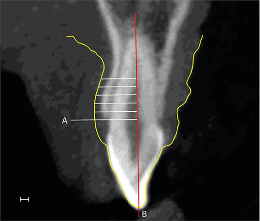
Figure 1 Para-axial slice at the mid-buccal aspect of lateral incisor. Outline of gingiva which is obtained from scanned file is marked as yellow line. Thickness measurements at 1–5 mm from the alveolar crest (A); and perpendicular to the root axis (B), bar=1 mm.
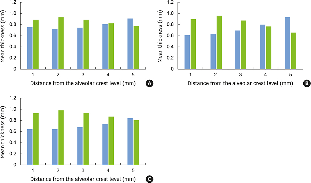
Figure 2 Mean thicknesses of hard and soft tissues in each level (1–5 mm under the alveolar crest level). Blue and green bars indicate gingival and buccal bone thicknesses, respectively. (A) Central incisors; (B) Lateral incisors; and (C) Canines.
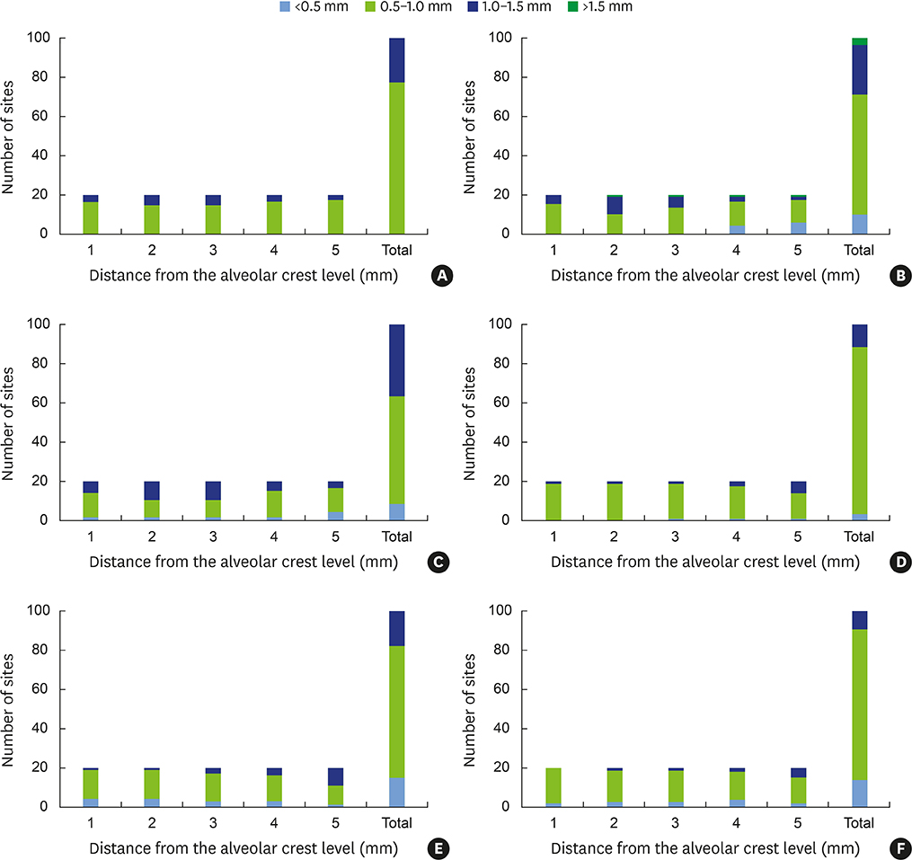
Figure 3 Frequency distribution of buccal bone and gingival thickness per point of measurement. (A) Frequency of buccal bone thickness at central incisors; (B) Frequency of buccal bone thickness at lateral incisors; (C) Frequency of buccal bone thickness at canines; (D) Frequency of gingival thickness at central incisors; (E) Frequency of gingival thickness at lateral incisors; and (F) Frequency of gingival thickness at canines.
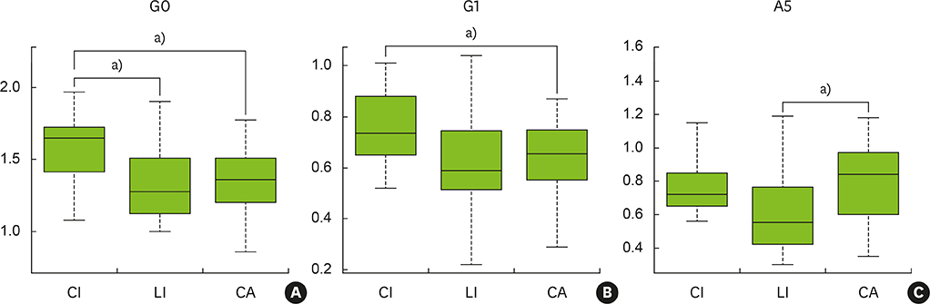
Figure 4 Comparison between buccal bone and gingival thicknesses according to tooth types. There was a significant difference among tooth types for G0, G1, and A5. CI, central incisor; LI, lateral incisor; CA, canine; G0, gingival thickness at alveolar crest line; G1, gingival thickness at 1 mm inferior to the alveolar crest; A5, buccal bone thickness at 5 mm inferior to the alveolar crest. a)Statistically significant difference (P<0.05).
Reference
-
1. Olsson M, Lindhe J. Periodontal characteristics in individuals with varying form of the upper central incisors. J Clin Periodontol. 1991; 18:78–82.
Article2. Weisgold AS. Contours of the full crown restoration. Alpha Omegan. 1977; 70:77–89.3. Evans CD, Chen ST. Esthetic outcomes of immediate implant placements. Clin Oral Implants Res. 2008; 19:73–80.
Article4. Kois JC. Predictable single-tooth peri-implant esthetics: five diagnostic keys. Compend Contin Educ Dent. 2004; 25:895–896.5. Hwang D, Wang HL. Flap thickness as a predictor of root coverage: a systematic review. J Periodontol. 2006; 77:1625–1634.
Article6. Baldi C, Pini-Prato G, Pagliaro U, Nieri M, Saletta D, Muzzi L, et al. Coronally advanced flap procedure for root coverage. Is flap thickness a relevant predictor to achieve root coverage? A 19-case series. J Periodontol. 1999; 70:1077–1084.
Article7. Fu JH, Yeh CY, Chan HL, Tatarakis N, Leong DJ, Wang HL. Tissue biotype and its relation to the underlying bone morphology. J Periodontol. 2010; 81:569–574.
Article8. Stein JM, Lintel-Höping N, Hammächer C, Kasaj A, Tamm M, Hanisch O. The gingival biotype: measurement of soft and hard tissue dimensions - a radiographic morphometric study. J Clin Periodontol. 2013; 40:1132–1139.
Article9. Botticelli D, Berglundh T, Lindhe J. Hard-tissue alterations following immediate implant placement in extraction sites. J Clin Periodontol. 2004; 31:820–828.
Article10. Lee SL, Kim HJ, Son MK, Chung CH. Anthropometric analysis of maxillary anterior buccal bone of Korean adults using cone-beam CT. J Adv Prosthodont. 2010; 2:92–96.
Article11. Januário AL, Duarte WR, Barriviera M, Mesti JC, Araújo MG, Lindhe J. Dimension of the facial bone wall in the anterior maxilla: a cone-beam computed tomography study. Clin Oral Implants Res. 2011; 22:1168–1171.
Article12. Kan JY, Rungcharassaeng K, Umezu K, Kois JC. Dimensions of peri-implant mucosa: an evaluation of maxillary anterior single implants in humans. J Periodontol. 2003; 74:557–562.
Article13. De Rouck T, Eghbali R, Collys K, De Bruyn H, Cosyn J. The gingival biotype revisited: transparency of the periodontal probe through the gingival margin as a method to discriminate thin from thick gingiva. J Clin Periodontol. 2009; 36:428–433.
Article14. Kan JY, Morimoto T, Rungcharassaeng K, Roe P, Smith DH. Gingival biotype assessment in the esthetic zone: visual versus direct measurement. Int J Periodontics Restorative Dent. 2010; 30:237–243.15. Claffey N, Shanley D. Relationship of gingival thickness and bleeding to loss of probing attachment in shallow sites following nonsurgical periodontal therapy. J Clin Periodontol. 1986; 13:654–657.
Article16. La Rocca AP, Alemany AS, Levi P Jr, Juan MV, Molina JN, Weisgold AS. Anterior maxillary and mandibular biotype: relationship between gingival thickness and width with respect to underlying bone thickness. Implant Dent. 2012; 21:507–515.17. Younes F, Eghbali A, Raes M, De Bruyckere T, Cosyn J, De Bruyn H. Relationship between buccal bone and gingival thickness revisited using non-invasive registration methods. Clin Oral Implants Res. 2016; 27:523–528.
Article18. Sanz Martin I, Benic GI, Hämmerle CH, Thoma DS. Prospective randomized controlled clinical study comparing two dental implant types: volumetric soft tissue changes at 1 year of loading. Clin Oral Implants Res. 2016; 27:406–411.
Article19. Windisch SI, Jung RE, Sailer I, Studer SP, Ender A, Hämmerle CH. A new optical method to evaluate three-dimensional volume changes of alveolar contours: a methodological in vitro study. Clin Oral Implants Res. 2007; 18:545–551.
Article20. Nikiforidou M, Tsalikis L, Angelopoulos C, Menexes G, Vouros I, Konstantinides A. Classification of periodontal biotypes with the use of CBCT. A cross-sectional study. Clin Oral Investig. 2016; 20:2061–2071.
Article21. Nowzari H, Molayem S, Chiu CH, Rich SK. Cone beam computed tomographic measurement of maxillary central incisors to determine prevalence of facial alveolar bone width ≥2 mm. Clin Implant Dent Relat Res. 2012; 14:595–602.
Article22. Müller HP, Schaller N, Eger T, Heinecke A. Thickness of masticatory mucosa. J Clin Periodontol. 2000; 27:431–436.
Article
- Full Text Links
-
- Actions
-
Cited
- CITED
-
- Close
- Share
-
- Similar articles
-
- Analysis of the root position and angulation of maxillary premolars in alveolar bone using cone-beam computed tomography
- Evaluation of mandibular cortical bone thickness for placement of temporary anchorage devices (TADs)
- Correlation analysis of periodontal tissue dimensions in the esthetic zone using a non-invasive digital method
- The effect of periodontal therapy on the gingival thickness in patients with drug-induced gingival enlargement
- Quantitative evaluation of cortical bone and soft tissue thickness in the mandible
