J Korean Ophthalmol Soc.
2016 Dec;57(12):1958-1963. 10.3341/jkos.2016.57.12.1958.
Posterior Surface Opacification of a Silicone Intraocular Lens in a Patient with Asteroid Hyalosis
- Affiliations
-
- 1Department of Ophthalmology, Soonchunhyang University College of Medicine, Cheonan, Korea. wismile@schmc.ac.kr
- 2SU Yonsei Eye Clinic, Seoul, Korea.
- KMID: 2362716
- DOI: http://doi.org/10.3341/jkos.2016.57.12.1958
Abstract
- PURPOSE
In the present study, a case of posterior surface opacification of a silicone intraocular lens (IOL) in a patient with asteroid hyalosis (AH) is reported.
CASE SUMMARY
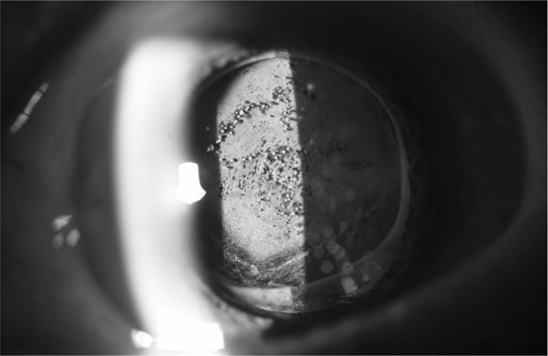
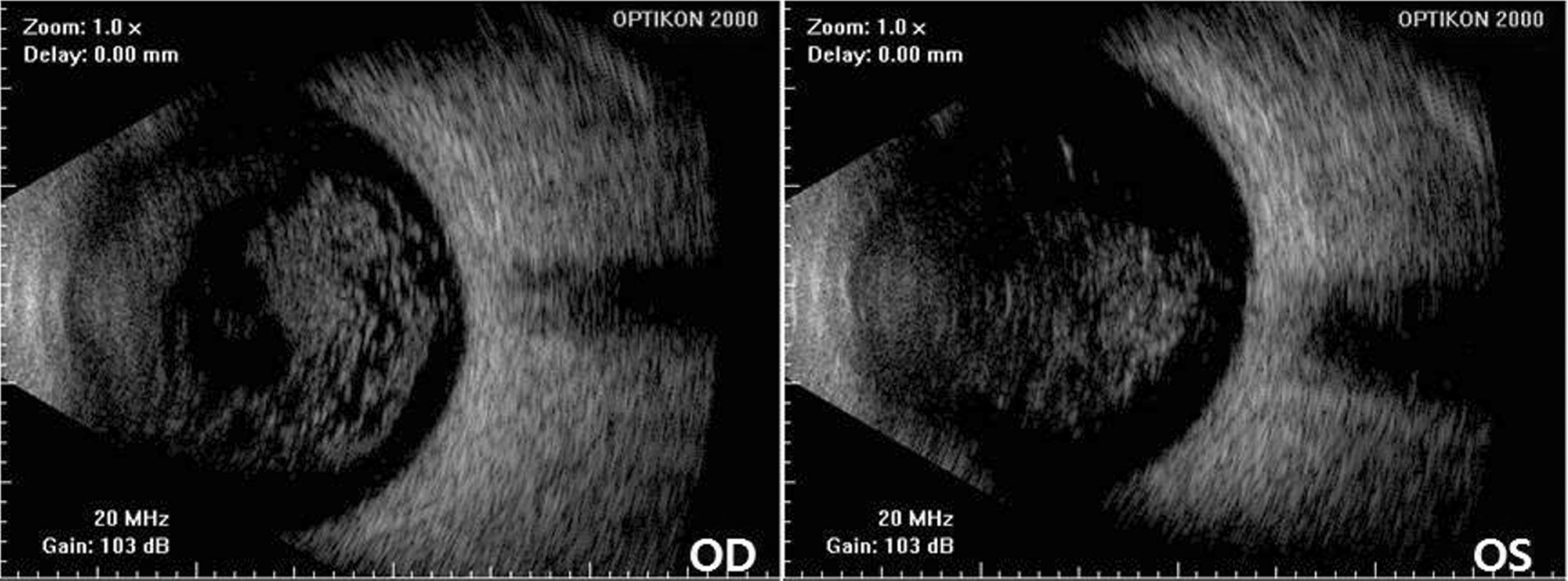
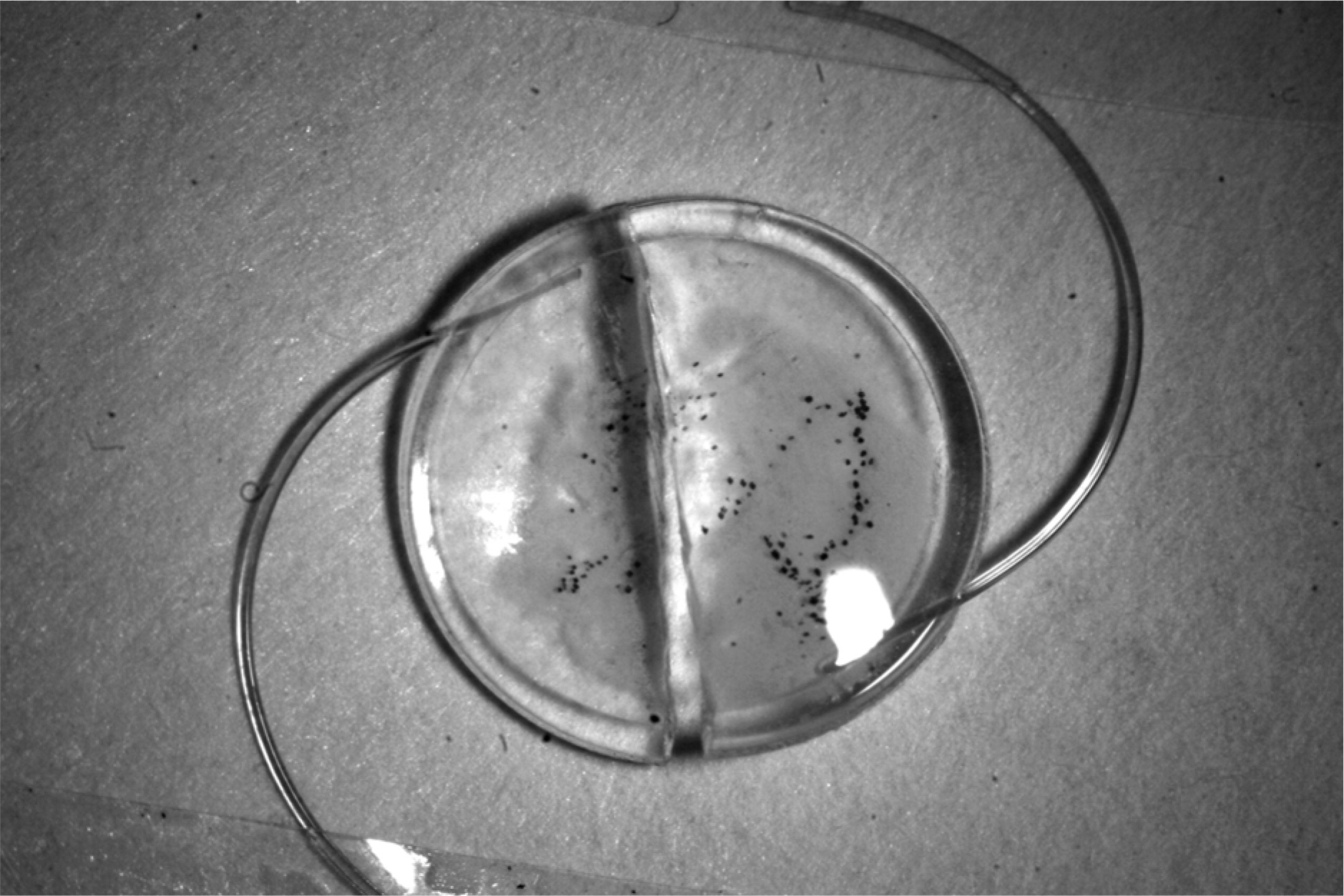
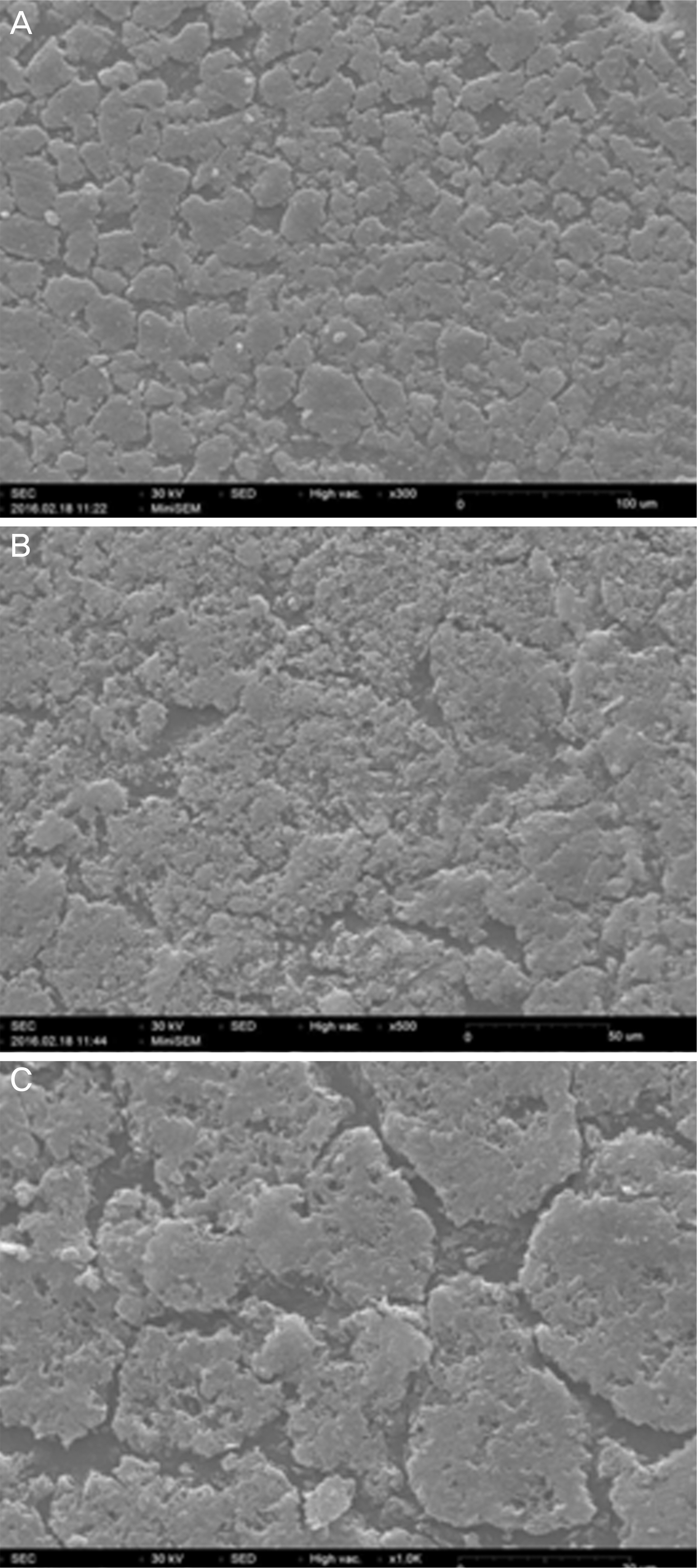
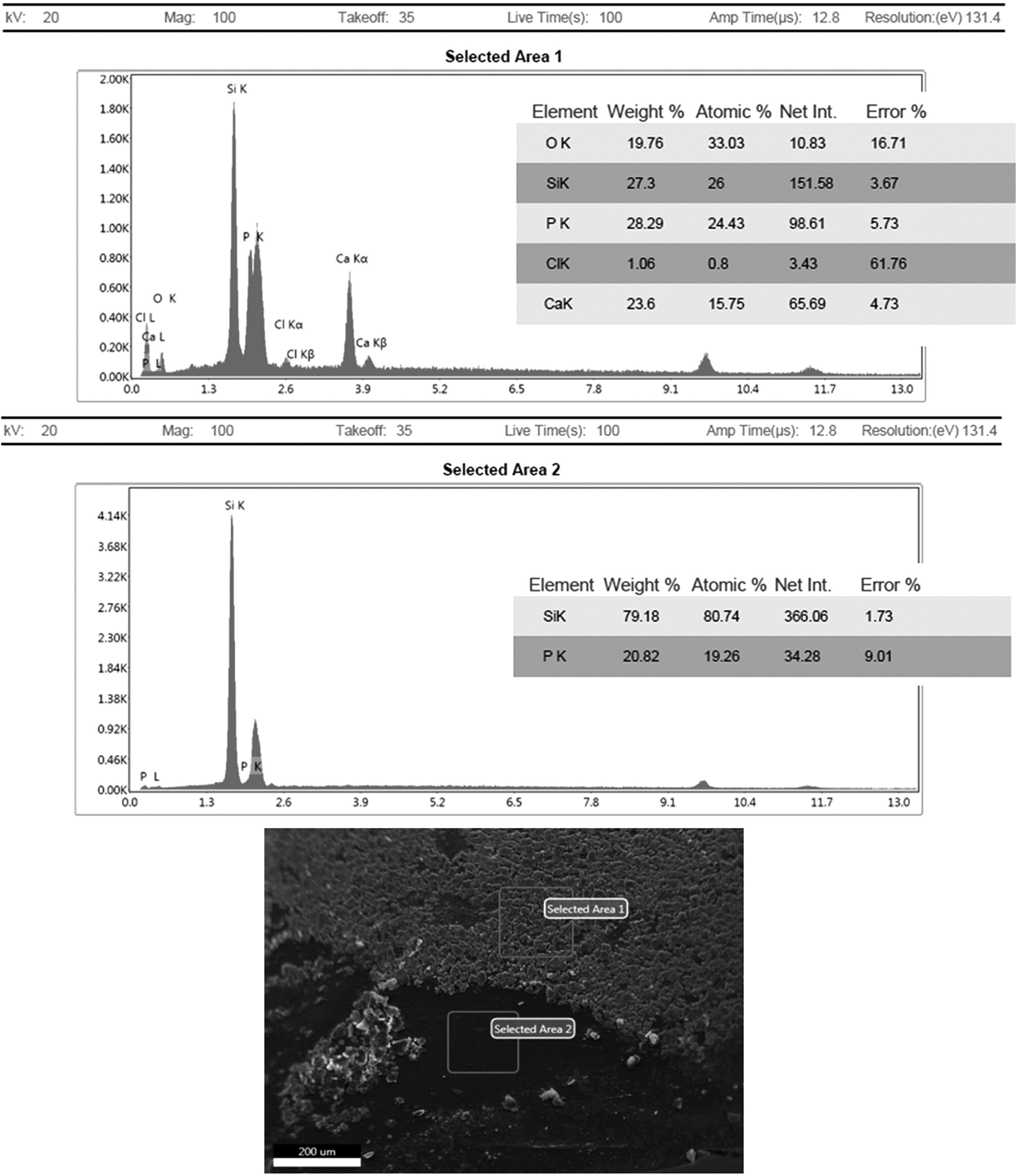
76-year-old male was referred to our clinic with IOL opacification in his left eye. The patient had uneventful cataract surgery 7 years prior with the same silicone IOL implanted in both eyes. Three years after surgery, posterior capsular opacity was observed in his left eye and neodymium:YAG (Nd:YAG) laser capulotomy was performed. After posterior capsulotomy, opacification of the IOL's posterior surface was observed on slit lamp examination. IOL exchange was performed and the explanted IOL was analyzed using a light microscope and a scanning electron microscope with energy dispersive X-ray spectroscopy for elemental analysis of the deposits. The calcification was on the posterior surface of the IOL and composed mainly of calcium and phosphorus, the main components of AH. The right eye showed clear IOL with intact posterior lens capsule.
CONCLUSIONS
Surgeons performing cataract surgery should consider the possibility of surface calcification of silicone IOLs in eyes with AH before IOL selection for implantation.
MeSH Terms
Figure
Reference
-
References
1. Lee JY, Joo KM, Kim SH. Late opacification of hydrophilic acrylic intraocular lenses. J Korean Ophthalmol Soc. 2002; 43:2419–29.2. Kim HG, Lee SH, Choi YJ. Late postoperative opacification of the foldable hydrophilic acrylic intraocular lens, ACRL-160. J Korean Ophthalmol Soc. 2003; 44:315–20.3. Kim JC, Kim CS, Choi SH, et al. Clinical characteristics of patients with opacification of hydrophilic acrylic intraocular lens after abdominal surgery. J Korean Ophthalmol Soc. 2005; 46:1281–90.4. Kim SH, Choi SH. Determine a proper axial length by ultrasonic biometry in opaque hydrophilic acrylic intraocular lens. J Korean Ophthalmol Soc. 2008; 49:1453–60.
Article5. Lee SJ, Choi JH, Sun HJ, et al. Surface calcification of hydrophilic acrylic intraocular lens related to inflammatory membrane abdominal after combined vitrectomy and cataract surgery. J Cataract Refract Surg. 2010; 36:676–81.6. Moon K, Kim KS, Kim YC. A case of hydrophilic acrylic abdominal lens opacification in a patient with proliferative diabetic retinopathy. J Korean Ophthalmol Soc. 2012; 53:1172–6.7. Mitchell P, Wang MY, Wang JJ. Asteroid hyalosis in an older abdominal: The Blue Mountains Eye Study. Ophthalmic Epidemiol. 2003; 10:331–5.8. Winkler J, Lünsdorf H. Ultrastructure and composition of asteroid bodies. Invest Ophthalmol Vis Sci. 2001; 42:902–7.9. Foot L, Werner L, Gills JP, et al. Surface calcification of silicone plate intraocular lenses in patients with asteroid hyalosis. Am J Ophthalmol. 2004; 137:979–87.10. Wackernagel W, Ettinger K, Weitgasser U, et al. Opacification of a silicone intraocular lens caused by calcium deposits on the optic. J Cataract Refract Surg. 2004; 30:517–20.
Article11. Werner L, Kollarits CR, Mamalis N, Olson RJ. Surface calcification of a 3-piece silicone intraocular lens in a patient with asteroid hyalosis: a clinicopathologic case report. Ophthalmology. 2005; 112:447–52.12. Stringham J, Werner L, Monson B, et al. Calcification of different designs of silicone intraocular lenses in eyes with asteroid hyalosis. Ophthalmology. 2010; 117:1486–92.
Article13. Matsumura K, Takano M, Shimizu K, Nemoto N. Silicone abdominal lens surface calcification in a patient with asteroid hyalosis. Jpn J Ophthalmol. 2012; 56:319–23.
- Full Text Links
- Actions
-
Cited
- CITED
-
- Close
- Share
- Similar articles
-
- Asteroid Hyalosis that Caused Decreased Vision after Cataract Surgery
- Clinical Manifestation of Asteroid Hyalosis
- Posterior Capsule Opacification and Intraocular Lens Design in Sulcus Fixated Posterior Chamber Lens
- Posterior Capsule Opacification and Intraocular Lens Design with In-the-bag fixated Posterior Chamber Lens
- Two Cases of Intraoperative Acute Opacification of Hydrophilic Intraocular Lens

