Clin Orthop Surg.
2015 Dec;7(4):490-496. 10.4055/cios.2015.7.4.490.
Arthroscopic Assessment of Intra-Articular Lesion after Surgery for Rotational Ankle Fracture
- Affiliations
-
- 1Department of Orthopedic Surgery, Saeum Hospital, Seoul, Korea.
- 2Department of Orthopedic Surgery, Maryknoll Hospital, Busan, Korea. jazzkari@nate.com
- 3Department of Orthopedic Surgery, Inje University Busan Paik Hospital, Inje University College of Medicine, Busan, Korea.
- KMID: 2360264
- DOI: http://doi.org/10.4055/cios.2015.7.4.490
Abstract
- BACKGROUND
The purpose of this study was to report findings of exploratory arthroscopic assessment performed in conjunction with removal of internal fixation device placed in the initial surgery for rotational ankle fracture.
METHODS
A total of 53 patients (33 male, 20 female) who underwent surgery for rotational ankle fracture between November 2002 and February 2008 were retrospectively reviewed. All patients gave consent to the exploratory arthroscopic surgery for the removal of internal fixation devices placed in the initial surgery. Lauge-Hansen classification system of ankle fractures was assessed for all patients. Intra-articular lesions (osteochondral lesion, loose body, and fibrosis) were evaluated via ankle arthroscopy. Comparative analysis was then performed between radiological classification of ankle fracture/patient's symptoms and arthroscopic findings.
RESULTS
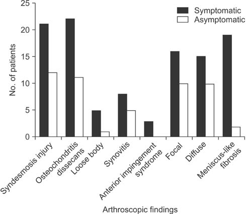
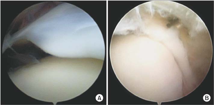
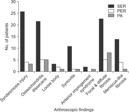
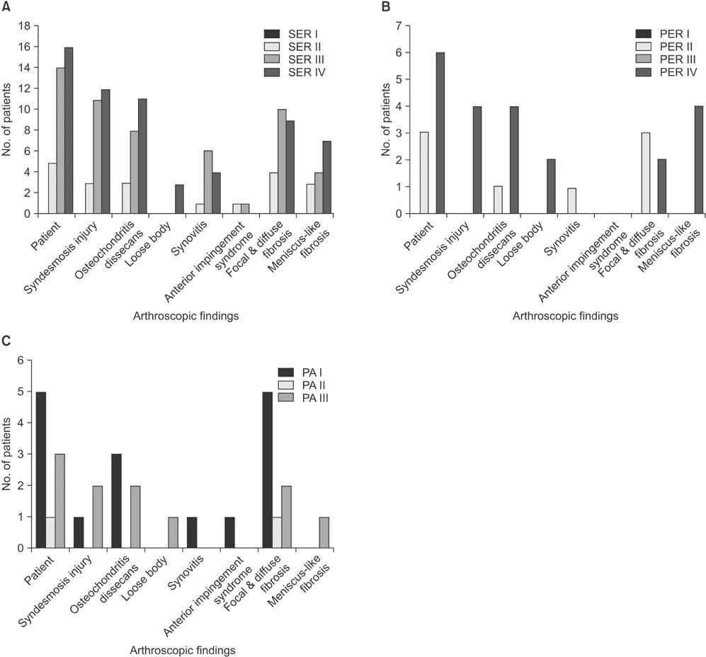
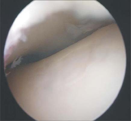
Lauge-Hansen classification system of ankle fractures included supination-external rotation type (n = 35), pronation-external rotation type (n = 9), and pronation-abduction type (n = 9). A total of 33 patients exhibited symptoms of pain or discomfort while walking whereas 20 exhibited no symptoms. Arthroscopic findings included abnormal findings around the syndesmosis area (n = 35), intra-articular fibrosis (n = 51), osteochondral lesions of the talus (n = 33), loose bodies (n = 6), synovitis (n = 13), and anterior bony impingement syndrome (n = 3). Intra-articular fibrosis was seen in 31 of symptomatic patients (93.9%). Pain or discomfort with activity caused by soft tissue impingement with meniscus-like intra-articular fibrosis were found in 19 patients. There was statistical significance (p = 0.02) between symptoms (pain and discomfort) and the findings of meniscus-like fibrosis compared to the group without any symptom.
CONCLUSIONS
Arthroscopic examination combined with treatment of intra-articular fibrosis arising from ankle fracture surgery may help improve surgical outcomes.
Keyword
MeSH Terms
Figure
Reference
-
1. Jaivin JS, Ferkel RD. Arthroscopy of the foot and ankle. Clin Sports Med. 1994; 13(4):761–783.
Article2. Thomas B, Yeo JM, Slater GL. Chronic pain after ankle fracture: an arthroscopic assessment case series. Foot Ankle Int. 2005; 26(12):1012–1016.
Article3. Utsugi K, Sakai H, Hiraoka H, Yashiki M, Mogi H. Intra-articular fibrous tissue formation following ankle fracture: the significance of arthroscopic debridement of fibrous tissue. Arthroscopy. 2007; 23(1):89–93.
Article4. Beris AE, Kabbani KT, Xenakis TA, Mitsionis G, Soucacos PK, Soucacos PN. Surgical treatment of malleolar fractures: a review of 144 patients. Clin Orthop Relat Res. 1997; (341):90–98.
Article5. Day GA, Swanson CE, Hulcombe BG. Operative treatment of ankle fractures: a minimum ten-year follow-up. Foot Ankle Int. 2001; 22(2):102–106.
Article6. Berndt AL, Harty M. Transchondral fractures (osteochondritis dissecans) of the talus. J Bone Joint Surg Am. 1959; 41(6):988–1020.
Article7. Ferkel RD, Scranton PE Jr. Arthroscopy of the ankle and foot. J Bone Joint Surg Am. 1993; 75(8):1233–1242.
Article8. Hintermann B, Regazzoni P, Lampert C, Stutz G, Gachter A. Arhroscopic findings in acute fractures of the ankle. J Bone Joint Surg Br. 2000; 82(3):345–351.9. Takao M, Uchio Y, Naito K, Fukazawa I, Kakimaru T, Ochi M. Diagnosis and treatment of combined intra-articular disorders in acute distal fibular fractures. J Trauma. 2004; 57(6):1303–1307.
Article10. Bonnin M, Bouysset M. Arthroscopy of the ankle: analysis of results and indications on a series of 75 cases. Foot Ankle Int. 1999; 20(11):744–751.
Article11. van Dijk CN, Bossuyt PM, Marti RK. Medial ankle pain after lateral ligament rupture. J Bone Joint Surg Br. 1996; 78(4):562–567.
Article12. Gulish HA, Sullivan RJ, Aronow M. Arthroscopic treatment of soft-tissue impingement lesions of the ankle in adolescents. Foot Ankle Int. 2005; 26(3):204–207.
Article13. Henderson I, La Valette D. Ankle impingement: combined anterior and posterior impingement syndrome of the ankle. Foot Ankle Int. 2004; 25(9):632–638.
Article14. Lee JW, Suh JS, Huh YM, Moon ES, Kim SJ. Soft tissue impingement syndrome of the ankle: diagnostic efficacy of MRI and clinical results after arthroscopic treatment. Foot Ankle Int. 2004; 25(12):896–902.
Article15. Russo A, Zappia M, Reginelli A, et al. Ankle impingement: a review of multimodality imaging approach. Musculoskelet Surg. 2013; 97:Suppl 2. S161–S168.
Article16. Linklater JM. Imaging of talar dome chondral and osteochondral lesions. Top Magn Reson Imaging. 2010; 21(1):3–13.
Article17. Loren GJ, Ferkel RD. Arthroscopic assessment of occult intra-articular injury in acute ankle fractures. Arthroscopy. 2002; 18(4):412–421.
Article18. Martin DF, Baker CL, Curl WW, Andrews JR, Robie DB, Haas AF. Operative ankle arthroscopy: long-term followup. Am J Sports Med. 1989; 17(1):16–23.19. Ogilvie-Harris DJ, Gilbart MK, Chorney K. Chronic pain following ankle sprains in athletes: the role of arthroscopic surgery. Arthroscopy. 1997; 13(5):564–574.
Article
- Full Text Links
- Actions
-
Cited
- CITED
-
- Close
- Share
- Similar articles
-
- The Effect of Temporary K-wire Fixation in the Plate Fixation for Displaced Intra-articular Calcaneal Fracture
- Arthroscopic Procedure in the Treatment of Chronic Lateral Ankle Instability
- Arthroscopic Treatment of Isolated Anterior Malleolar Fracture of the Ankle
- Arthroscopic Assessment of Potential Intra-articular Ankle Injury in Treatment of Ankle Fracture
- Treatment of Articular Fracture of the Distal Tibia (Pilon Fracture) with Limited Open Reduction and External Fixator

