J Korean Foot Ankle Soc.
2015 Dec;19(4):151-155. 10.14193/jkfas.2015.19.4.151.
Arthroscopic Assessment of Potential Intra-articular Ankle Injury in Treatment of Ankle Fracture
- Affiliations
-
- 1Department of Orthopedic Surgery, Busan Paik Hospital, Inje University College of Medicine, Busan, Korea. ortho1@hanmail.net
- 2Departmnt of Medicine, Recruit Training Center of the 8th Army Division, Republic of Korea Army, Korea.
- KMID: 2132456
- DOI: http://doi.org/10.14193/jkfas.2015.19.4.151
Abstract
- PURPOSE
The purpose of this study was to analyze the frequency and patterns of intra-articular lesions detected during ankle fracture surgery using ankle arthroscopy.
MATERIALS AND METHODS
Thirty patients (31 ankles) who underwent open reduction and internal fixation combined with ankle arthroscopy for acute ankle fracture at Inje University Busan Paik Hospital from June 2011 to September 2013 were evaluated. The ankle fractures were classified according to the AO/OTA (AO Foundation and Orthopaedic Trauma Association) classification and the intraarticular injuries were identified by ankle arthroscopy. Osteochondral lesions of the talus were divided into nine subtypes based on their locations, and the ligament injuries were classified according to avulsion fracture and rupture.
RESULTS
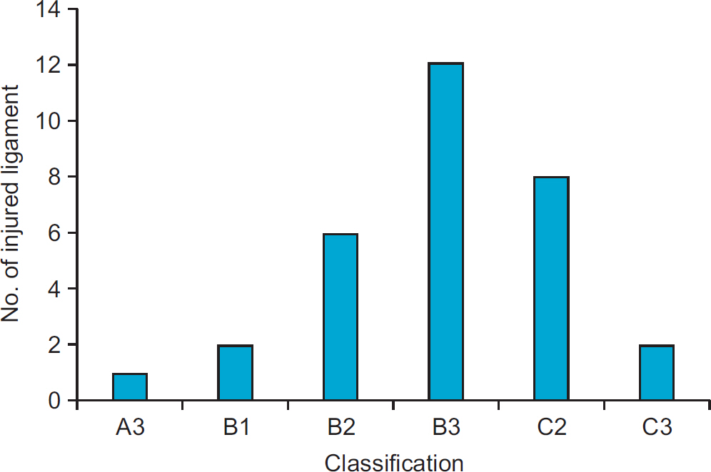
Using arthroscopy, abnormality in the distal tibiofibular ligament was found in 21 cases and osteochondral lesions and defects of the talus larger than 5 mm were detected in 26 cases. Among ligament injuries, anterior inferior tibio-fibular ligament injury was found in 14 cases, posterior inferior tibio-fibular ligament injury was found in two cases, deep deltoid ligament injury was found in three cases, and deep transverse tibio-fibular ligament injury was found in five cases. The locations of the osteochondral lesions were on the antero-lateral, antero-medial, centro-medial, centro-central, centro-lateral, and postero-lateral talus in 11, one, two, one, two, and nine cases, respectively.
CONCLUSION
With early diagnosis and treatment arthroscopy performed at the time of intra-articular fracture surgery is expected to result in a good outcome.
Keyword
MeSH Terms
Figure
Reference
-
References
1. Loren GJ, Ferkel RD. Arthroscopic assessment of occult intraarticular injury in acute ankle fractures. Arthroscopy. 2002; 18:412–21.
Article2. Thomas B, Yeo JM, Slater GL. Chronic pain after ankle fracture: an arthroscopic assessment case series. Foot Ankle Int. 2005; 26:1012–6.
Article3. Thordarson DB, Bains R, Shepherd LE. The role of ankle arthroscopy on the surgical management of ankle fractures. Foot Ankle Int1. 2001; 22:123–5.
Article4. Harper MC. Ankle fracture classification systems: a case for integration of the Lauge-Hansen and AO-Danis-Weber schemes. Foot Ankle. 1992; 13:404–7.
Article5. Lauge-Hansen N. Ligamentous ankle fractures; diagnosis and treatment. Acta Chir Scand. 1949; 97:544–50.6. de Leeuw PA, van Sterkenburg MN, van Dijk CN. Arthroscopy and endoscopy of the ankle and hindfoot. Sports Med Arthrosc. 2009; 17:175–84.
Article7. Sung KS. Ankle arthroscopy: anatomy, portals and instrument. J Korean Foot Ankle Soc. 2012; 16:1–8.8. Chu IT, Kim YS, Yoo SH, Oh IS. The incidences and locations of osteochondral lesions of the talus in ankle fracture. J Korean Orthop Assoc. 2004; 39:494–7.
Article9. Boraiah S, Paul O, Parker RJ, Miller AN, Hentel KD, Lorich DG. Osteochondral lesions of talus associated with ankle fractures. Foot Ankle Int. 2009; 30:481–5.
Article10. Hintermann B, Regazzoni P, Lampert C, Stutz G, Gächter A. Arthroscopic findings in acute fractures of the ankle. J Bone Joint Surg Br. 2000; 82:345–51.
Article11. Stufkens SA, Knupp M, Horisberger M, Lampert C, Hintermann B. Cartilage lesions and the development of osteoarthritis after internal fixation of ankle fractures: a prospective study. J Bone Joint Surg Am. 2010; 92:279–86.12. Taga I, Shino K, Inoue M, Nakata K, Maeda A. Articular cartilage lesions in ankles with lateral ligament injury. An arthroscopic study. Am J Sports Med. 1993; 21:120–7.13. Bauer M, Bergström B, Hemborg A, Sandegård J. Malleolar fractures: nonoperative versus operative treatment. A controlled study. Clin Orthop Relat Res. 1985; 199:17–27.14. Cedell CA. Supination-outward rotation injuries of the ankle: a clinical and roentgenological study with special reference to the operative treatment. Acta Orthop Scand. 1967; 38(Suppl 110):1–148.
Article15. Hughes JL, Weber H, Willenegger H, Kuner EH. Evaluation of ankle fractures: nonoperative and operative treatment. Clin Orthop Relat Res. 1979; 138:111–9.16. Klossner O. Late results of operative and nonoperative treatment of severe ankle fractures. A clinical study. Acta Chir Scand Suppl. 1962; (Suppl 293):1–93.17. Choi GW, Choi WJ, Lee JW. Ankle impingement syndrome. J Korean Foot Ankle Soc. 2012; 16:19–25.18. Sorrento DL, Mlodzienski A. Incidence of lateral talar dome lesions in SER IV ankle fractures. J Foot Ankle Surg. 2000; 39:354–8.
Article19. Takao M, Ochi M, Naito K, Uchio Y, Kono T, Oae K. Arthroscopic drilling for chondral, subchondral, and combined chondral-subchondral lesions of the talar dome. Arthroscopy. 2003; 19:524–30.
Article20. Utsugi K, Sakai H, Hiraoka H, Yashiki M, Mogi H. Intra-articular fibrous tissue formation following ankle fracture: the significance of arthroscopic debridement of fibrous tissue. Arthros-copy. 2007; 23:89–93.
Article
- Full Text Links
- Actions
-
Cited
- CITED
-
- Close
- Share
- Similar articles
-
- Arthroscopic Procedure in the Treatment of Chronic Lateral Ankle Instability
- Intra-Articular Hyaluronic Acid Injection in Ankle Osteoarthritis
- Arthroscopic Treatment of Isolated Anterior Malleolar Fracture of the Ankle
- Arthroscopic Assessment of Intra-Articular Lesion after Surgery for Rotational Ankle Fracture
- Arthroscopic Modified Broström Operation for Lateral Ankle Instability


