Total Lesion Glycolysis Using 18F-FDG PET/CT as a Prognostic Factor for Locally Advanced Esophageal Cancer
- Affiliations
-
- 1Department of Internal Medicine, Incheon St. Mary's Hospital, Incheon, Korea.
- 2Department of Internal Medicine, Seoul St. Mary's Hospital, Seoul, Korea.
- 3Department of Nuclear Medicine, Daejeon St. Mary's Hospital, Daejeon, Korea.
- 4Department of Radiation Oncology, Seoul St. Mary's Hospital, Seoul, Korea.
- 5Department of Nuclear Medicine, Incheon St. Mary's Hospital, Incheon, Korea.
- 6Department of Nuclear Medicine, Seoul St. Mary's Hospital, The Catholic University of Korea, Seoul, Korea. iryoo@catholic.ac.kr
- KMID: 2359990
- DOI: http://doi.org/10.3346/jkms.2016.31.1.39
Abstract
- Standardized uptake value (SUV), metabolic tumor volume (MTV), and total lesion glycolysis (TLG) have been considered prognostic factors for survival in many cancers. However, their prognostic value for radiotherapy-treated squamous esophageal cancer has not been evaluated. In this study, SUV, MTV, and TLG were measured to predict their prognostic role in overall survival (OS) in 38 esophageal cancer patients who had undergone 18F-FDG PET/CT before radiotherapy. TLG demonstrated higher sensitivity and specificity for predicting OS than MTV and SUV; and a better OS was observed in patients with low TLG compared to those with high TLG in locally advanced disease (OS, 46.9 months; 95% confidence interval [CI], 33.50-60.26 vs. 25.3 months; 95% CI, 8.37-42.28; P=0.003). Multivariate analyses in these patients determined that TLG and the use of combination chemotherapy were the independent prognostic factors for OS (hazard ratio [HR], 7.12; 95% CI, 2.038-24.857; P=0.002 and HR, 6.76; 95% CI, 2.149-21.248; P=0.001, respectively). These results suggest that TLG is an independent prognostic factor for OS and a better predictor of survival than MTV and SUV in patients with locally advanced esophageal cancer treated with radiotherapy.
MeSH Terms
-
Adult
Aged
Aged, 80 and over
Area Under Curve
Esophageal Neoplasms/mortality/pathology/*radiography
Female
Fluorodeoxyglucose F18/chemistry
Glycolysis/*physiology
Humans
Male
Middle Aged
Neoplasm Staging
*Positron-Emission Tomography
Prognosis
Proportional Hazards Models
ROC Curve
Radiopharmaceuticals/*chemistry
Retrospective Studies
Survival Rate
*Tomography, X-Ray Computed
Fluorodeoxyglucose F18
Radiopharmaceuticals
Figure
-

Fig. 1 Receiver operating characteristic curve analysis of survival prediction according to metabolic parameters in the 38 patients (continuous variable). Area under the curve of total lesion glycolysis (TLG), metabolic tumor volume (MTV), mean standardized uptake value (SUVmean) were 0.695 (P=0.041, 95% confidence interval [CI], 0.522-0.867), 0.678 (P=0.046, 95% CI, 0.507-0.820), and 0.609 (P=0.252, 95% CI, 0.428-0.791), respectively. The differences in the AUC between TLG and MTV, TLG and SUVmean, and MTV and SUVmean were 0.017 (P=0.559), 0.086 (P=0.266), and 0.069 (P=0.460), respectively. Thus, the optimal cut-off values of TLG and MTV were determined as 232.98 and 44.7. Sensitivity and specificity of the dichotomized TLG (≤232.98 vs. >232.98) and MTV (≤44.7 vs. >44.7) were 71.4/76.5% and 61.9/82.35%, respectively.

Fig. 2 Overall survival. (A) Total patients; Upper line, T1,T2 (n = 18; 40.8 months, 95% confidence interval [CI], 21.56-60.10) and lower line, T3, T4 (n = 15; 32.3 months, 95% CI, 20.46-44.20). P = 0.385. (B) Patients with locally advanced esophageal cancer; Upper line, TLG ≤ 232.98 (n = 14; 46.9 months, 95% CI, 33.50-60.26) and lower line, TLG > 232.98 (n = 19; 25.3 months, 95% CI, 8.37-42.28). P = 0.003. (C) Patients with locally advanced esophageal cancer; Upper line, MTV ≤ 44.70 cm3 (n = 17; 39.0 months, 95% CI, 26.37-51.63) and lower line, MTV > 44.70 cm3 (n = 16; 34.6 months, 95,% CI 15.10-54.01). P = 0.054. (D) Patients with locally advanced esophageal cancer; Upper line, SUVmean ≤ 5.70 (n = 16; 43.0 months, 95% CI, 21.11-64.80) and lower line, SUVmean > 5.70 (n = 17; 31.0 months, 95% CI, 17.10-44.89). P = 0.963.
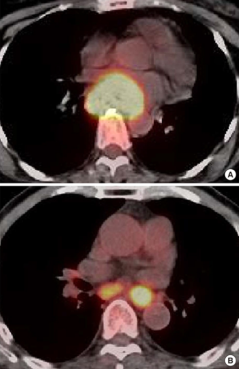
Fig. 3 Representative examples of the relationship between metabolic parameters and overall survival. (A) Representative 18F-FDG PET/CT images of a 52-yr-old female patient with middle esophageal cancer (T3N0, stage IIB) exhibiting a TLG of 673.92, an MTV of 93.6 cm3, and a SUVmax of 21.7. She exhibited progressive disease after 5-fluorouracil/cisplatin chemoradiotherapy and lived for 5.6 months after the diagnosis. (B) Representative 18F-FDG PET/CT images of a 79-yr-old male patient with lower esophageal cancer (T2N1, stage IIB) exhibiting a TLG of 68.64, an MTV of 15.6 cm3, and a SUVmax of 10.5. He exhibited stable disease after radiotherapy alone and lived for 61.8 months after the diagnosis.
Reference
-
1. Parkin DM, Bray F, Ferlay J, Pisani P. Global cancer statistics, 2002. CA Cancer J Clin. 2005; 55:74–108.2. Stahl M, Stuschke M, Lehmann N, Meyer HJ, Walz MK, Seeber S, Klump B, Budach W, Teichmann R, Schmitt M, et al. Chemoradiation with and without surgery in patients with locally advanced squamous cell carcinoma of the esophagus. J Clin Oncol. 2005; 23:2310–2317.3. Chan SC, Chang JT, Lin CY, Ng SH, Wang HM, Liao CT, Chang CJ, Lin SY, Yen TC. Clinical utility of 18F-FDG PET parameters in patients with advanced nasopharyngeal carcinoma: predictive role for different survival endpoints and impact on prognostic stratification. Nucl Med Commun. 2011; 32:989–996.4. Lee HY, Hyun SH, Lee KS, Kim BT, Kim J, Shim YM, Ahn MJ, Kim TS, Yi CA, Chung MJ. Volume-based parameter of 18)F-FDG PET/CT in malignant pleural mesothelioma: prediction of therapeutic response and prognostic implications. Ann Surg Oncol. 2010; 17:2787–2794.5. Chung HH, Kwon HW, Kang KW, Park NH, Song YS, Chung JK, Kang SB, Kim JW. Prognostic value of preoperative metabolic tumor volume and total lesion glycolysis in patients with epithelial ovarian cancer. Ann Surg Oncol. 2012; 19:1966–1972.6. Dibble EH, Alvarez AC, Truong MT, Mercier G, Cook EF, Subramaniam RM. 18F-FDG metabolic tumor volume and total glycolytic activity of oral cavity and oropharyngeal squamous cell cancer: adding value to clinical staging. J Nucl Med. 2012; 53:709–715.7. Liao S, Penney BC, Wroblewski K, Zhang H, Simon CA, Kampalath R, Shih MC, Shimada N, Chen S, Salgia R, et al. Prognostic value of metabolic tumor burden on 18F-FDG PET in nonsurgical patients with non-small cell lung cancer. Eur J Nucl Med Mol Imaging. 2012; 39:27–38.8. Eisenhauer EA, Therasse P, Bogaerts J, Schwartz LH, Sargent D, Ford R, Dancey J, Arbuck S, Gwyther S, Mooney M, et al. New response evaluation criteria in solid tumours: revised RECIST guideline (version 1.1). Eur J Cancer. 2009; 45:228–247.9. Zhang J, Jia Z, Ragaz J, Zhang YJ, Zhou M, Zhang YP, Li G, Wang BY, Wang ZH, Hu XC. The maximum standardized uptake value of 18 F-FDG PET scan to determine prognosis of hormone-receptor positive metastatic breast cancer. BMC Cancer. 2013; 13:42.10. Lin SC, Liao CY, Kao CH, Yen KY, Yang SN, Wang YC, Liang JA, Chen SW. Pretreatment maximal standardized uptake value of the primary tumor predicts outcome to radiotherapy in patients with pharyngeal cancer. J Radiat Res (Tokyo). 2012; 53:462–468.11. Davies A, Tan C, Paschalides C, Barrington SF, O'Doherty M, Utley M, Treasure T. FDG-PET maximum standardised uptake value is associated with variation in survival: analysis of 498 lung cancer patients. Lung Cancer. 2007; 55:75–78.12. Atsumi K, Nakamura K, Abe K, Hirakawa M, Shioyama Y, Sasaki T, Baba S, Isoda T, Ohga S, Yoshitake T, et al. Prediction of outcome with FDG-PET in definitive chemoradiotherapy for esophageal cancer. J Radiat Res (Tokyo). 2013; 54:890–898.13. Palie O, Michel P, Ménard JF, Rousseau C, Rio E, Bridji B, Benyoucef A, Meyer ME, Jalali K, Bardet S, et al. The predictive value of treatment response using FDG PET performed on day 21 of chemoradiotherapy in patients with oesophageal squamous cell carcinoma. A prospective, multicentre study (RTEP3). Eur J Nucl Med Mol Imaging. 2013; 40:1345–1355.14. Song SY, Kim JH, Ryu JS, Lee GH, Kim SB, Park SI, Song HY, Cho KJ, Ahn SD, Lee SW, et al. FDG-PET in the prediction of pathologic response after neoadjuvant chemoradiotherapy in locally advanced, resectable esophageal cancer. Int J Radiat Oncol Biol Phys. 2005; 63:1053–1059.15. Hyun SH, Choi JY, Shim YM, Kim K, Lee SJ, Cho YS, Lee JY, Lee KH, Kim BT. Prognostic value of metabolic tumor volume measured by 18F-fluorodeoxyglucose positron emission tomography in patients with esophageal carcinoma. Ann Surg Oncol. 2010; 17:115–122.16. Choi ES, Ha SG, Kim HS, Ha JH, Paeng JC, Han I. Total lesion glycolysis by 18F-FDG PET/CT is a reliable predictor of prognosis in soft-tissue sarcoma. Eur J Nucl Med Mol Imaging. 2013; 40:1836–1842.17. Moon SH, Hyun SH, Choi JY. Prognostic significance of volume-based PET parameters in cancer patients. Korean J Radiol. 2013; 14:1–12.18. Chen HH, Chiu NT, Su WC, Guo HR, Lee BF. Prognostic value of whole-body total lesion glycolysis at pretreatment FDG PET/CT in non-small cell lung cancer. Radiology. 2012; 264:559–566.19. Arslan N, Tuncel M, Kuzhan O, Alagoz E, Budakoglu B, Ozet A, Ozguven MA. Evaluation of outcome prediction and disease extension by quantitative 2-deoxy-2-[18F] fluoro-D-glucose with positron emission tomography in patients with small cell lung cancer. Ann Nucl Med. 2011; 25:406–413.20. Moon SH, Choi JY, Lee HJ, Son YI, Baek CH, Ahn YC, Park K, Lee KH, Kim BT. Prognostic value of 18F-FDG PET/CT in patients with squamous cell carcinoma of the tonsil: comparisons of volume-based metabolic parameters. Head Neck. 2013; 35:15–22.21. Yoo J, Choi JY, Lee KT, Heo JS, Park SB, Moon SH, Choe YS, Lee KH, Kim BT. Prognostic significance of volume-based metabolic parameters by (18)F-FDG PET/CT in gallbladder carcinoma. Nucl Med Mol Imaging. 2012; 46:201–206.22. Hatt M, Le Pogam A, Visvikis D, Pradier O, Cheze Le Rest C. Impact of partial-volume effect correction on the predictive and prognostic value of baseline 18F-FDG PET images in esophageal cancer. J Nucl Med. 2012; 53:12–20.23. Li YM, Lin Q, Zhao L, Wang LC, Sun L, Dai MM, Luo ZM, Zheng H, Wu H. Pre-treatment metabolic tumor volume and total lesion glycolysis are useful prognostic factors for esophageal squamous cell cancer patients. Asian Pac J Cancer Prev. 2014; 15:1369–1373.24. Aquino SL, Halpern EF, Kuester LB, Fischman AJ. FDG-PET and CT features of non-small cell lung cancer based on tumor type. Int J Mol Med. 2007; 19:495–499.
- Full Text Links
-
- Actions
-
Cited
- CITED
-
- Close
- Share
-
- Similar articles
-
- Use of 18F-FDG PET/CT in Second Primary Cancer
- Pretreatment 18F‑FDG PET/CT‑Derived Parameters in Predicting Clinical Outcomes of Locally Advanced Upper Third Esophageal Squamous Cell Carcinoma After Definitive Chemoradiation Therapy
- Significance of Metabolic Tumor Volume and Total Lesion Glycolysis Measured Using ¹â¸F-FDG PET/CT in Locally Advanced and Metastatic Gallbladder Carcinoma
- Esophageal Leiomyoma with intense FDG uptake on 18F-FDG PET/CT
- Preoperative Nodal ¹â¸F-FDG Avidity Rather than Primary Tumor Avidity Determines the Prognosis of Patients with Advanced Gastric Cancer
