Yonsei Med J.
2019 Jul;60(7):604-610. 10.3349/ymj.2019.60.7.604.
Significance of Metabolic Tumor Volume and Total Lesion Glycolysis Measured Using ¹â¸F-FDG PET/CT in Locally Advanced and Metastatic Gallbladder Carcinoma
- Affiliations
-
- 1Division of Medical Oncology, Department of Internal Medicine, Yonsei Cancer Center, Yonsei University College of Medicine, Seoul, Korea.
- 2Division of Medical Oncology, Department of Internal Medicine, Gangnam Severance Hospital, Yonsei University College of Medicine, Seoul, Korea.
- 3Division of Medical Oncology, Department of Internal Medicine, St. Vincent's Hospital, The Catholic University of Korea, Suwon, Korea.
- 4Cancer Prevention Center, Yonsei Cancer Center, Yonsei University College of Medicine, Seoul, Korea.
- 5Department of Nuclear Medicine, Gangnam Severance Hospital, Yonsei University College of Medicine, Seoul, Korea. tjeonnm@yuhs.ac
- KMID: 2450399
- DOI: http://doi.org/10.3349/ymj.2019.60.7.604
Abstract
- PURPOSE
This study aimed to determine the prognostic value of new quantitative parameters of 18F-fluorodeoxyglucose positron emission tomography/computed tomography (18F-FDG PET/CT), including metabolic tumor volume (MTV), in patients with locally advanced and metastatic gallbladder cancer (GBC).
MATERIALS AND METHODS
In total, 83 patients initially diagnosed with locally advanced and metastatic GBC and who underwent 18F-FDG PET/CT at the time of initial diagnosis were retrospectively reviewed. The metabolic volume-based PET parameters of primary tumors and metastatic lesions were measured, including maximum and average standardized uptake values (SUV), MTV, and total lesion glycolysis. An overall survival (OS) analysis was performed using the Kaplan-Meier method with PET and clinical parameters. A Cox proportional hazards regression analysis was performed to determine independent prognostic factors.
RESULTS
In univariate analysis, pathologic differentiation (p<0.001), performance status (PS; p=0.003), C-reactive protein (CRP) level (p=0.009), and PET-related SUVmt max (the highest SUV among the metastatic lesions) (p=0.040) and MTVtotal (the sum of the MTVs of both the primary and metastatic lesions) (p=0.031), were significant for OS. In multivariate analysis, MTVtotal (hazard ratio: 2.07; 95% confidence interval: 1.23-3.48; p=0.006) remained significant for the prediction of OS, as did differentiation (p=0.001), PS (p=0.001), and CRP (p=0.039).
CONCLUSION
In locally advanced and metastatic GBC, volume-based PET/CT parameters of the total tumor burden of malignancy, such as MTVtotal, were found to be useful for the identification of patients with poor prognosis.
MeSH Terms
Figure
-
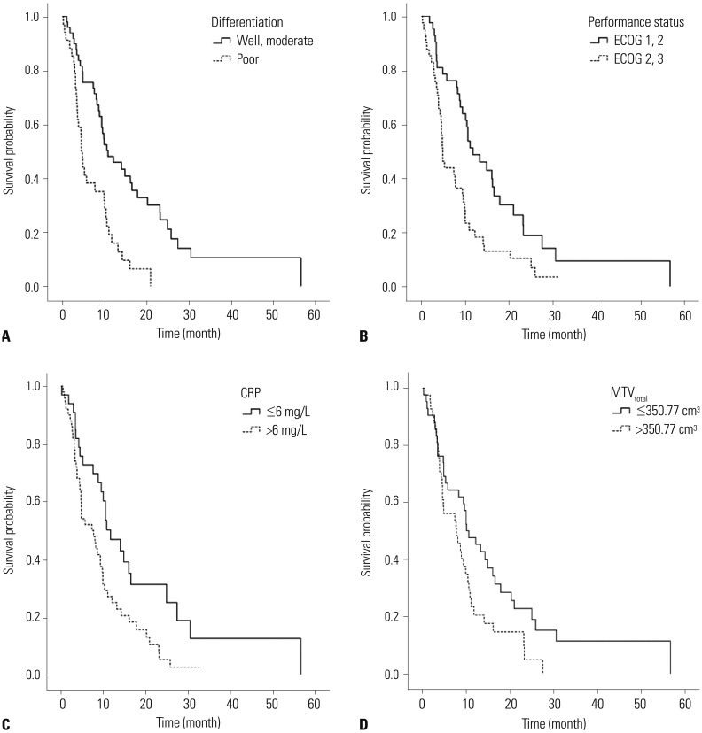
Fig. 1 Kaplan-Meier curves for overall survival based on significant prognostic factors, including (A) pathologic differentiation [HR=2.42 (well differentiated and moderately differentiated vs. poorly differentiated); p=0.001], (B) performance status [HR=2.28 (ECOG 0, 1 vs. 2, 3); p=0.001], (C) CRP [HR=1.73 (≤6 mg/dL vs. >6 mg/dL); p=0.039], and (D) MTVtotal [HR=2.07 (≤350.77 cm3 vs. >350.77 cm3); p=0.006] in gallbladder carcinoma. ECOG, Eastern Cooperative Oncology Group; CRP, C-reactive protein; MTVtotal, the sum of the MTVs of both the locally advanced and metastatic lesions; HR, hazard ratio.
Reference
-
1. Strom BL, Soloway RD, Rios-Dalenz JL, Rodriguez-Martinez HA, West SL, Kinman JL, et al. Risk factors for gallbladder cancer. An international collaborative case-control study. Cancer. 1995; 76:1747–1756. PMID: 8625043.
Article2. Randi G, Franceschi S, La Vecchia C. Gallbladder cancer worldwide: geographical distribution and risk factors. Int J Cancer. 2006; 118:1591–1602. PMID: 16397865.
Article3. Corvera CU, Blumgart LH, Akhurst T, DeMatteo RP, D'Angelica M, Fong Y, et al. 18F-fluorodeoxyglucose positron emission tomography influences management decisions in patients with biliary cancer. J Am Coll Surg. 2008; 206:57–65. PMID: 18155569.
Article4. Allal AS, Dulguerov P, Allaoua M, Haenggeli CA, El-Ghazi el A, Lehmann W, et al. Standardized uptake value of 2-[(18)F] fluoro-2-deoxy-D-glucose in predicting outcome in head and neck carcinomas treated by radiotherapy with or without chemotherapy. J Clin Oncol. 2002; 20:1398–1404. PMID: 11870185.
Article5. Davies A, Tan C, Paschalides C, Barrington SF, O'Doherty M, Utley M, et al. FDG-PET maximum standardised uptake value is associated with variation in survival: analysis of 498 lung cancer patients. Lung Cancer. 2007; 55:75–78. PMID: 17084485.
Article6. Hyun SH, Choi JY, Shim YM, Kim K, Lee SJ, Cho YS, et al. Prognostic value of metabolic tumor volume measured by 18F-fluorodeoxyglucose positron emission tomography in patients with esophageal carcinoma. Ann Surg Oncol. 2010; 17:115–122. PMID: 19826877.
Article7. Lee HY, Hyun SH, Lee KS, Kim BT, Kim J, Shim YM, et al. Volume-based parameter of 18F-FDG PET/CT in malignant pleural mesothelioma: prediction of therapeutic response and prognostic implications. Ann Surg Oncol. 2010; 17:2787–2794. PMID: 20461469.8. Moon SH, Choi JY, Lee HJ, Son YI, Baek CH, Ahn YC, et al. Prognostic value of 18F-FDG PET/CT in patients with squamous cell carcinoma of the tonsil: comparisons of volume-based metabolic parameters. Head Neck. 2013; 35:15–22. PMID: 22307893.9. Lee P, Weerasuriya DK, Lavori PW, Quon A, Hara W, Maxim PG, et al. Metabolic tumor burden predicts for disease progression and death in lung cancer. Int J Radiat Oncol Biol Phys. 2007; 69:328–333. PMID: 17869659.
Article10. Ciernik IF, Dizendorf E, Baumert BG, Reiner B, Burger C, Davis JB, et al. Radiation treatment planning with an integrated positron emission and computer tomography (PET/CT): a feasibility study. Int J Radiat Oncol Biol Phys. 2003; 57:853–863. PMID: 14529793.
Article11. Ramos-Font C, Gómez-Rio M, Rodríguez-Fernández A, Jiménez-Heffernan A, Sánchez Sánchez R, Llamas-Elvira JM. Ability of FDG-PET/CT in the detection of gallbladder cancer. J Surg Oncol. 2014; 109:218–224. PMID: 24165875.
Article12. Bos R, van Der Hoeven JJ, van Der Wall E, van Der Groep P, van Diest PJ, Comans EF, et al. Biologic correlates of (18)fluorodeoxyglucose uptake in human breast cancer measured by positron emission tomography. J Clin Oncol. 2002; 20:379–387. PMID: 11786564.
Article13. Kurokawa T, Yoshida Y, Kawahara K, Tsuchida T, Okazawa H, Fujibayashi Y, et al. Expression of GLUT-1 glucose transfer, cellular proliferation activity and grade of tumor correlate with [F-18]-fluorodeoxyglucose uptake by positron emission tomography in epithelial tumors of the ovary. Int J Cancer. 2004; 109:926–932. PMID: 15027127.
Article14. Chung JK, Lee YJ, Kim SK, Jeong JM, Lee DS, Lee MC. Comparison of [18F]fluorodeoxyglucose uptake with glucose transporter-1 expression and proliferation rate in human glioma and non-small-cell lung cancer. Nucl Med Commun. 2004; 25:11–17. PMID: 15061260.
Article15. Donohue JH. Present status of the diagnosis and treatment of gallbladder carcinoma. J Hepatobiliary Pancreat Surg. 2001; 8:530–534. PMID: 11956904.16. Donohue JH, Stewart AK, Menck HR. The National Cancer Data Base report on carcinoma of the gallbladder, 1989–1995. Cancer. 1998; 83:2618–2628. PMID: 9874470.
Article17. Manfredi S, Benhamiche AM, Isambert N, Prost P, Jouve JL, Faivre J. Trends in incidence and management of gallbladder carcinoma: a population-based study in France. Cancer. 2000; 89:757–762. PMID: 10951337.18. Furukawa H, Ikuma H, Asakura K, Uesaka K. Prognostic importance of standardized uptake value on F-18 fluorodeoxyglucose-positron emission tomography in biliary tract carcinoma. J Surg Oncol. 2009; 100:494–499. PMID: 19653260.
Article19. Yoo J, Choi JY, Lee KT, Heo JS, Park SB, Moon SH, et al. Prognostic significance of volume-based metabolic parameters by (18)F-FDG PET/CT in gallbladder carcinoma. Nucl Med Mol Imaging. 2012; 46:201–206. PMID: 24900061.
Article20. Maldonado A, González-Alenda FJ, Alonso M, Sierra JM. PET-CT in clinical oncology. Clin Transl Oncol. 2007; 9:494–505. PMID: 17720652.
Article21. Fendler WP, Philippe Tiega DB, Ilhan H, Paprottka PM, Heinemann V, Jakobs TF, et al. Validation of several SUV-based parameters derived from 18F-FDG PET for prediction of survival after SIRT of hepatic metastases from colorectal cancer. J Nucl Med. 2013; 54:1202–1208. PMID: 23729697.
Article
- Full Text Links
-
- Actions
-
Cited
- CITED
-
- Close
- Share
-
- Similar articles
-
- Prognostic Significance of Volume-based Metabolic Parameters by 18F-FDG PET/CT in Gallbladder Carcinoma
- Prognostic Value of Metabolic Information in Advanced Gastric Cancer Using Preoperative ¹â¸F-FDG PET/CT
- Prognostic value of metabolic tumor volume and total lesion glycolysis from ¹â¸F-FDG PET/CT in lymph node metastases and risk stratification of endometrial carcinoma
- Focal Nasopharyngeal Activity Detected on [18F]FDG PET/CT: Clinical Implications and Comparison of Metabolic Parameters for Prediction of Malignancy
- ¹â¸F-FDG PET/MR Refines Evaluation in Newly Diagnosed Metastatic Urethral Adenocarcinoma
