J Korean Assoc Oral Maxillofac Surg.
2016 Oct;42(5):307-314. 10.5125/jkaoms.2016.42.5.307.
Retiform hemangioendothelioma in the infratemporal fossa and buccal area: a case report and literature review
- Affiliations
-
- 1Division of Oral and Maxillofacial Surgery, Department of Dentistry, Inha University School of Medicine, Incheon, Korea. kik@inha.ac.kr
- 2Department of Oral and Maxillofacial Surgery, Catholic Kwandong University International St. Mary's Hospital, Incheon, Korea.
- KMID: 2356259
- DOI: http://doi.org/10.5125/jkaoms.2016.42.5.307
Abstract
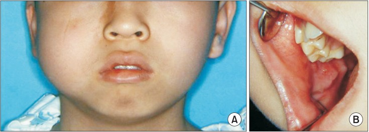
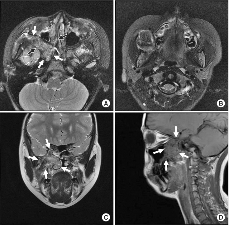
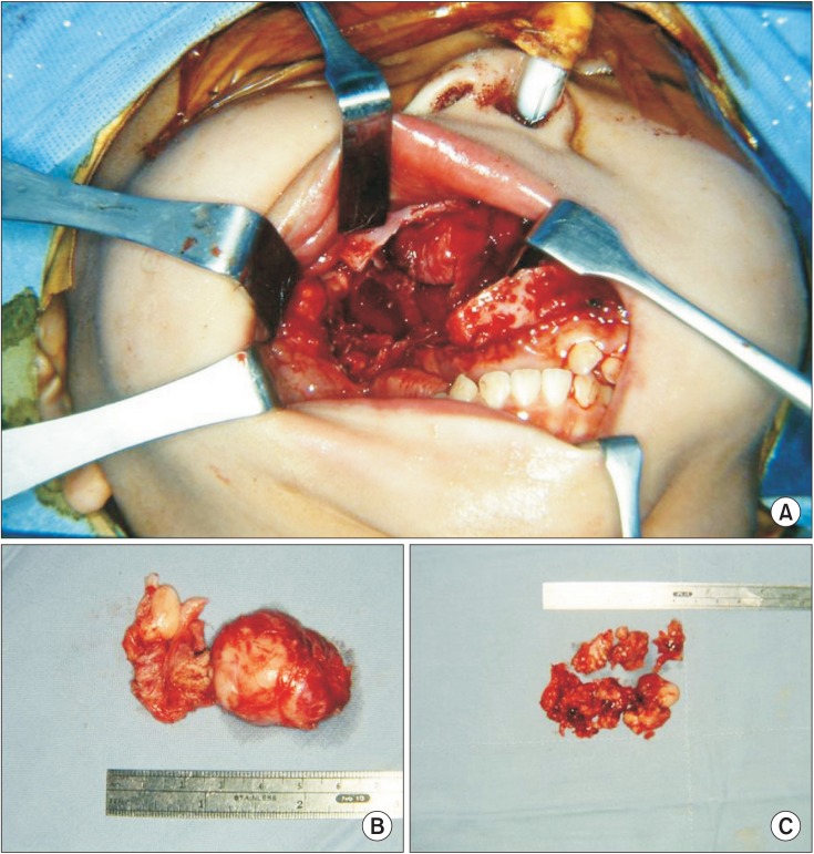
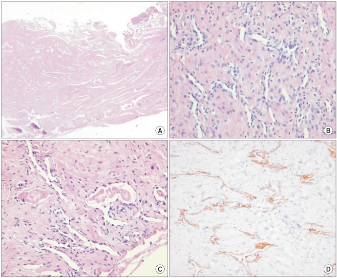
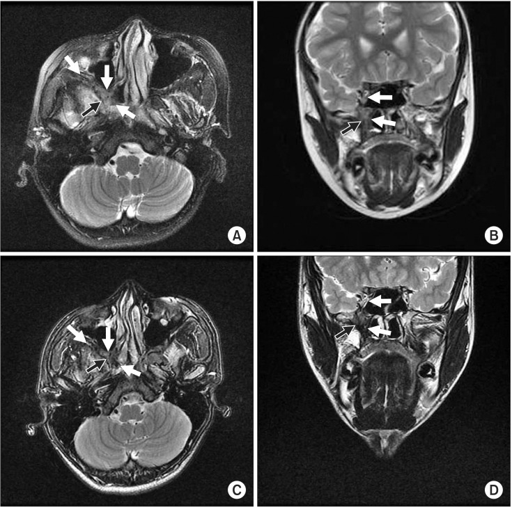
- We report a case of retiform hemangioendothelioma (RH) located in the infratemporal fossa and buccal area in a 13-year-old Korean boy. The tumor originated from the sphenoid bone of the infratemporal fossa area and spread into the cavernous sinus, orbital apex, and retro-nasal area with bone destruction of the pterygoid process. Tumor resection was conducted via Le Fort I osteotomy and partial maxillectomy to approach the infratemporal fossa and retro-nasal area. The diagnosis of RH was confirmed after surgery. In the presented patient, surgical excision was incomplete, and close follow-up was performed. There was no evidence of expansion or metastasis of the residual tumor in the 8 years after surgery. In cases of residual RH with low likelihood of expansion and metastasis, even though RH is an intermediate malignancy, close follow-up can be the appropriate treatment choice over additional aggressive therapy. To date, 29 papers and 48 RH cases have been reported, including this case. This case is the second reported RH case presenting as primary bone tumor and the first case originating in the oromaxillofacial area.
MeSH Terms
Figure
Reference
-
1. Calonje E, Fletcher CD, Wilson-Jones E, Rosai J. Retiform hemangioendothelioma. A distinctive form of low-grade angiosarcoma delineated in a series of 15 cases. Am J Surg Pathol. 1994; 18:115–125. PMID: 8291650.2. Fukunaga M, Endo Y, Masui F, Yoshikawa T, Ishikawa E, Ushigome S. Retiform haemangioendothelioma. Virchows Arch. 1996; 428:301–304. PMID: 8764941.
Article3. Aditya GS, Santosh V, Yasha TC, Shankar SK. Epithelioid and retiform hemangioendothelioma of the skull bone--report of four cases. Indian J Pathol Microbiol. 2003; 46:645–649. PMID: 15025366.4. El Darouti M, Marzouk SA, Sobhi RM, Bassiouni DA. Retiform hemangioendothelioma. Int J Dermatol. 2000; 39:365–368. PMID: 10849129.
Article5. Tan D, Kraybill W, Cheney RT, Khoury T. Retiform hemangioendothelioma: a case report and review of the literature. J Cutan Pathol. 2005; 32:634–637. PMID: 16176302.
Article6. Serel S, Serel BI, Uluc A, Heper AO, Gultan MS. Congenital retiform hemangioendothelioma. Indian J Dermatol. 2007; 52:160–162.
Article7. O'Duffy F, Timon C, Toner M. A rare angiosarcoma: retiform haemangioendothelioma. J Laryngol Otol. 2012; 126:200–202. PMID: 21888747.8. Albertini AF, Brousse N, Bodemer C, Calonje E, Fraitag S. Retiform hemangioendothelioma developed on the site of an earlier cystic lymphangioma in a six-year-old girl. Am J Dermatopathol. 2011; 33:e84–e87. PMID: 21915027.
Article9. Zhang G, Lu Q, Yin H, Wen H, Su Y, Li D, et al. A case of retiform-hemangioendothelioma with unusual presentation and aggressive clinical features. Int J Clin Exp Pathol. 2010; 3:528–533. PMID: 20606734.10. Hirsh AZ, Yan W, Wei L, Wernicke AG, Parashar B. Unresectable retiform hemangioendothelioma treated with external beam radiation therapy and chemotherapy: a case report and review of the literature. Sarcoma. 2010; DOI: 10.1155/2010/756246.
Article11. Duke D, Dvorak A, Harris TJ, Cohen LM. Multiple retiform hemangioendotheliomas. A low-grade angiosarcoma. Am J Dermatopathol. 1996; 18:606–610. PMID: 8989934.12. Dufau JP, Pierre C, De Saint Maur PP, Bellavoir A, Gros P. Retiform hemangioendothelioma. Ann Pathol. 1997; 17:47–51. PMID: 9162159.13. Mentzel T, Stengel B, Katenkamp D. Retiform hemangioendothelioma. Clinico-pathologic case report and discussion of the group of low malignancy vascular tumors. Pathologe. 1997; 18:390–394. PMID: 9432675.14. Sanz-Trelles A, Rodrigo-Fernandez I, Ayala-Carbonero A, Contreras-Rubio F. Retiform hemangioendothelioma. A new case in a child with diffuse endovascular papillary endothelial proliferation. J Cutan Pathol. 1997; 24:440–444. PMID: 9274963.
Article15. Schommer M, Herbst RA, Brodersen JP, Kiehl P, Katenkamp D, Kapp A, et al. Retiform hemangioendothelioma: another tumor associated with human herpesvirus type 8? J Am Acad Dermatol. 2000; 42:290–292. PMID: 10642690.
Article16. Ulrich D, Hrynyschyn K. Fast growing multifocal retiform hemangioendothelioma: a case report. Eur J Plast Surg. 2002; 25:47–49.17. Coutinho MLF. Laser indications hemangiomas in children. An Acad Nac Med. 2002; 162:74–76.18. Escudero AG, Sanchez JS, Bustos GN, Serrano TG, Martin JJR, Campora RG. Retiform hemangioendothelioma: a report of two cases and literature review. Acta Dermosifiliogr. 2003; 94:102–106.19. Botros MF, Abdo I, Foda S, Amer H, Attia N, Saad S, et al. Retiform hemangioendothelioma. Egypt Dermatol Online J. 2005; 1:1–4.20. Ioannidou D, Panayiotides J, Krasagakis K, Stefanidou M, Manios A, Tosca A. Retiform hemangioendothelioma presenting as bruiselike plaque in an adult woman. Int J Dermatol. 2006; 45:53–55. PMID: 16426378.
Article21. Parsons A, Sheehan DJ, Sangueza OP. Retiform hemangioendotheliomas usually do not express D2-40 and VEGFR-3. Am J Dermatopathol. 2008; 30:31–33. PMID: 18212541.
Article22. Love WE, Keiler SA, Tamburro JE, Honda K, Gosain AK, Bordeaux JS. Surgical management of congenital dermatofibrosarcoma protuberans. J Am Acad Dermatol. 2009; 61:1014–1023. PMID: 19925926.
Article23. Bhutoria B, Konar A, Chakrabarti S, Das S. Retiform hemangioendothelioma with lymph node metastasis: a rare entity. Indian J Dermatol Venereol Leprol. 2009; 75:60–62. PMID: 19172034.
Article24. Emberger M, Laimer M, Steiner H, Zelger B. Retiform hemangioendothelioma: presentation of a case expressing D2-40. J Cutan Pathol. 2009; 36:987–990. PMID: 19674202.
Article25. Kajo K, Macháleková K, Pauer M. Retiform hemangioendotelioma in a 8-year-old girl--case report. Cesk Patol. 2009; 45:72–74. PMID: 19764161.26. Aydingöz IE, Mansur AT, Celasun B. Correspondence: retiform hemangioendothelioma presenting as a hyperhidrotic tumor. Int J Dermatol. 2010; 49:1076–1077. PMID: 20883275.27. Choi WK, Lee SH, Oh SA, Kang DH. Retiform hemangioendothelioma on the finger. Arch Plast Surg. 2012; 39:80–82. PMID: 22783501.
Article28. Findikçioğlu K, Findikçioğlu F, Kutlugün C, Karabağli P. Retiform hemangioendothelioma originated from capillary malformation: a rare case. Turk J Plast Surg. 2012; 20:12–15.29. Requena L, Kutzner H. Hemangioendothelioma. Semin Diagn Pathol. 2013; 30:29–44. PMID: 23327728.
Article
- Full Text Links
- Actions
-
Cited
- CITED
-
- Close
- Share
- Similar articles
-
- A Case of Endoscopic Drainage of Pterygoid Fossa Abscess Induced by Fungal Invasion
- A Case of Hemangiopericytoma Occurring in the Infratemporal Fossa
- A Case of Odontogenic Infratemporal Fossa Abscess
- A Case of Synovial Sarcoma Involved in the Infratemporal Fossa Resected with Subtemporal-Infratemporal Fossa Approach with Hearing Preservation
- A Study on the Variations in Arteries and Nerves of the Infratemporal fossa in Korean

