J Cerebrovasc Endovasc Neurosurg.
2016 Sep;18(3):271-275. 10.7461/jcen.2016.18.3.271.
Evaluation and Treatment of the Acute Cerebral Infarction with Convexal Subarachnoid Hemorrhage
- Affiliations
-
- 1Department of Neurosurgery, St. Vincent's Hospital, College of Medicine, The Catholic University of Korea, Suwon, Korea. tkddnr79@daum.net
- KMID: 2355653
- DOI: http://doi.org/10.7461/jcen.2016.18.3.271
Abstract
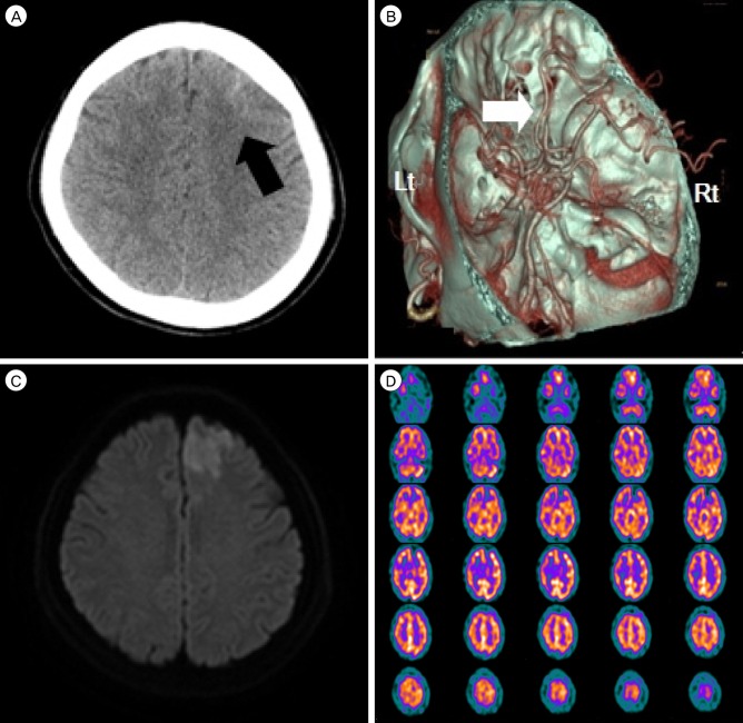
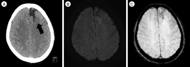
- Non-traumatic convexal subarachnoid hemorrhage (CSAH) is a comparatively infrequent with various vascular and nonvascular causes, it rarely occurs concomitant to acute ischemic stroke. We report a case of a 59-year-old woman, visited emergency room with right side subjective weakness spontaneously. Magnetic resonance diffusion-weighted images revealed an acute infarction of anterior cerebral arterial territory. Computed tomographic angiography showed a left frontal CSAH without any vascular lesions. And other laboratory studies were non-specific. We treated with dual antiplatelet drugs (cilostazole [Otsuka Pharmaceutical Co., Ltd. tokyo, Japan] and Aspirin [Bayer Pharma AG., Leverkusen, Germany]). She has done well for a follow-up period. (5 months) This case demonstrates the CSAH with acute infarction is rare but need to work up to identify the etiology and antiplatelet dugs are taken into account for treatments.
Keyword
MeSH Terms
Figure
Reference
-
1. Arévalo-Lorido JC, Carretero-Gómez J. Cerebral venous thrombosis with subarachnoid hemorrhage: a case report. Clin Med Res. 2015; 3. 13(1):40–43. PMID: 25380613.2. Arboix A, García-Eroles L, Sellarés N, Raga A, Oliveres M, Massons J. Infarction in the territory of the anterior cerebral artery: clinical study of 51 patients. BMC Neurol. 2009; 7. 9:30. PMID: 19589132.
Article3. Cuvinciuc V, Viguier A, Calviere L, Raposo N, Larrue V, Cognard C, et al. Isolated acute nontraumatic cortical subarachnoid hemorrhage. AJNR Am J Neuroradiol. 2010; 9. 31(8):1355–1362. PMID: 20093311.
Article4. Ducros A, Boukobza M, Porcher R, Sarov M, Valade D, Bousser MG. The clinical and radiological spectrum of reversible cerebral vasoconstriction syndrome. A prospective series of 67 patients. Brain. 2007; 12. 130(Pt 12):3091–3101. PMID: 18025032.
Article5. Finelli PF. Cerebral amyloid angiopathy as cause of convexity SAH in elderly. Neurologist. 2010; 1. 16(1):37–40. PMID: 20065795.
Article6. Geraldes R, Santos C, Canhão P. Atraumatic localized convexity subarachnoid hemorrhage associated with acute carotid artery occlusion. Eur J Neurol. 2011; 2. 18(2):e28–e29. PMID: 20868466.
Article7. Hentschel S, Toyota B. Intracranial malignant glioma presenting as subarachnoid hemorrhage. Can J Neurol Sci. 2003; 2. 30(1):63–66. PMID: 12619787.
Article8. Ito H, Uehara K, Matsumoto Y, Hashimoto A, Nagano C, Niimi M, et al. Cilostazol inhibits accumulation of triglyceride in aorta and platelet aggregation in cholesterol-fed rabbits. PLoS One. 2012; 7(6):e39374. PMID: 22761774.
Article9. Kang SY, Kim JS. Anterior cerebral artery infarction: stroke mechanism and clinical-imaging study in 100 patients. Neurology. 2008; 6. 70(24 Pt 2):2386–2393. PMID: 18541871.
Article10. Kannoth S, Iyer R, Thomas SV, Furtado SV, Rajesh BJ, Kesavadas C, et al. Intracranial infectious aneurysm: presentation, management and outcome. J Neurol Sci. 2007; 5. 256(1-2):3–9. PMID: 17360002.
Article11. Kazui S, Sawada T, Naritomi H, Kuriyama Y, Yamaguchi T. Angiographic evaluation of brain infarction limited to the anterior cerebral artery territory. Stroke. 1993; 4. 24(4):549–553. PMID: 8465361.
Article12. Kumar R, Wijdicks EF, Brown RD Jr, Parisi JE, Hammond CA. Isolated angiitis of the CNS presenting as subarachnoid haemorrhage. J Neurol Neurosurg Psychiatry. 1997; 6. 62(6):649–651. PMID: 9219758.
Article13. Kumar S, Goddeau RP Jr, Selim MH, Thomas A, Schlaug G, Alhazzani A, et al. Atraumatic convexal subarachnoid hemorrhage: clinical presentation, imaging patterns, and etiologies. Neurology. 2010; 3. 74(11):893–899. PMID: 20231664.14. Nakajima M, Inatomi Y, Yonehara T, Hirano T, Ando Y. Nontraumatic convexal subarachnoid hemorrhage concomitant with acute ischemic stroke. J Stroke Cerebrovasc Dis. 2014; 7. 23(6):1564–1570. PMID: 24630829.
Article15. Nakamura K, Ikomi F, Ohhashi T. Cilostazol, an inhibitor of type 3 phosphodiesterase, produces endothelium-independent vasodilation in pressurized rabbit cerebral penetrating arterioles. J Vasc Res. 2006; 43(1):86–94. PMID: 16286783.
Article16. Nonaka Y, Tsuruma K, Shimazawa M, Yoshimura S, Iwama T, Hara H. Cilostazol protects against hemorrhagic transformation in mice transient focal cerebral ischemia-induced brain damage. Neurosci Lett. 2009; 3. 452(2):156–161. PMID: 19383431.
Article17. O'Donnell MJ, Hankey GJ, Eikelboom JW. Antiplatelet therapy for secondary prevention of noncardioembolic ischemic stroke: a critical review. Stroke. 2008; 5. 39(5):1638–1646. PMID: 18369175.18. Rhode V, van Oosterhout A, Mull M, Gilsbach JM. Subarachnoid haemorrhage as initial symptom of multiple brain abscesses. Acta Neurochir (Wien). 2000; 142(2):205–208. PMID: 10795896.19. Sudo T, Tachibana K, Toga K, Tochizawa S, Inoue Y, Kimura Y, et al. Potent effects of novel anti-platelet aggregatory cilostamide analogues on recombinant cyclic nucleotide phosphodiesterase isozyme activity. Biochem Pharmacol. 2000; 2. 59(4):347–356. PMID: 10644042.
Article20. Sugiura Y, Morikawa T, Takenouchi T, Suematsu M, Kajimura M. Cilostazol strengthens the endothelial barrier of postcapillary venules from the rat mesentery in situ. Phlebology. 2014; 10. 29(9):594–599. PMID: 23858026.
Article21. Tan L, Margaret B, Zhang JH, Hu R, Yin Y, Cao L, et al. Efficacy and Safety of Cilostazol Therapy in Ischemic Stroke: A Meta-analysis. J Stroke Cerebrovasc Dis. 2015; 5. 24(5):930–938. PMID: 25804574.
Article22. Wan M, Han C, Xian P, Yang WZ, Li DS, Duan L. Moyamoya disease presenting with subarachnoid hemorrhage: Clinical features and neuroimaging of a case series. Br J Neurosurg. 2015; 29(6):804–810. PMID: 26313681.
Article23. Wang Y, Pan Y, Zhao X, Li H, Wang D, Johnston SC, et al. Clopidogrel With Aspirin in Acute Minor Stroke or Transient Ischemic Attack (CHANCE) Trial: One-Year Outcomes. Circulation. 2015; 7. 132(1):40–46. PMID: 25957224.
- Full Text Links
- Actions
-
Cited
- CITED
-
- Close
- Share
- Similar articles
-
- Simultaneous Nonaneurysmal Subarachnoid Hemorrhage and Acute Cerebral Infarction in a Patient with Intracranial Atherosclerosis
- Anesthetic Management of Cerebral Subarachnoid Hemorrhage with Intraoperative Electrocardiographic Change Simulating Acute Myocardial Infarction: A case report
- Terson Syndrome after Subarachnoid Hemorrhage Occurred by Thrombolysis and Mechanical Thrombectomy to Treat Acute Ischemic Stroke: A Case Report
- Progressive Manifestations of Reversible Cerebral Vasoconstriction Syndrome Presenting with Subarachnoid Hemorrhage, Intracerebral Hemorrhage, and Cerebral Infarction
- Isolated Acute Nontraumatic Cortical Subarachnoid Hemorrhage: Etiologies Based on MRI Findings


