The Diagnostic Dilemma of Neurolymphomatosis
- Affiliations
-
- 1Department of Neurology, Postgraduate Institute of Medical Education and Research (PGIMER), Chandigarh, India. goyal_mk@yahoo.com
- 2Department of Histopathology, Postgraduate Institute of Medical Education and Research (PGIMER), Chandigarh, India.
- 3Department of Radiodiagnosis, Postgraduate Institute of Medical Education and Research (PGIMER), Chandigarh, India.
- 4Department of Nuclear Medicine, Postgraduate Institute of Medical Education and Research (PGIMER), Chandigarh, India.
- 5Department of Clinical Hematology, Postgraduate Institute of Medical Education and Research (PGIMER), Chandigarh, India.
- KMID: 2354113
- DOI: http://doi.org/10.3988/jcn.2016.12.3.274
Abstract
- Neurolymphomatosis (NL) defined as infiltration of the central nervous system or the peripheral nervous system (PNS) by malignant lymphoma cells is a rare clinical entity. However, the increasing use of fluorodeoxyglucose positron-emission tomography (FDG-PET) and magnetic resonance imaging in evaluating PNS disorders is resulting in; this condition being recognized more frequently. Here; we report five NL patients and review the current literature. We report five patients with non-Hodgkin's lymphoma (NHL) and NL, all of whom were men aged 47-69 years. The clinical presentation varied from symmetrical peripheral neuropathy to mononeuropathy. Peripheral neuropathy was the presenting manifestation of a systemic lymphoma in two patients (40%). Neuroimaging as well as whole-body FDG-PET helped in determining the correct diagnosis in all of the patients. NL is an unusual presentation of NHL resulting from infiltration of the PNS by malignant lymphomatous cells. While evaluating peripheral neuropathy, a high degree of suspicion of NL is required since the presenting symptoms vary, conventional radiology has only modest sensitivity, and a pathological diagnosis is often difficult. FDG-PET helps in the early diagnosis and treatment of this condition.
Keyword
MeSH Terms
Figure
-
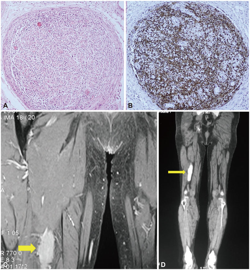
Fig. 1 Nerve bundle infiltration (patient 1), FDG-PET and MRI of right sciatic nerve (patient 2). A: A nerve bundle with infiltration by lymphoma cells (patient 1). B: CD 20 stain highlights diffuse infiltration by diffuse large B cells (patient 1). C: PET CT showing intense FDG uptake in right sciatic nerve (yellow arrow) (patient 2). D: intense enhancement of right sciatic nerve (arrow) on T1 weighted contrast MRI (patient 2).

Fig. 2 FDG-PET images (patient 4) and MRI Brain images (patient 5). A: FDG-PET showing intense uptake (white arrow) in the left testis (patient 4). B and C: Thickened and enhancing trigeminal nerves (yellow arrows) on fluid-attenuated inversion recovery and T1-weighted MRI, respectively (patient 5). FDG-PET: fluorodeoxyglucose positron tomography, MRI: magnetic resonance imaging.

Fig. 3 Nerve Biopsy staining (patient 4). A: Hematoxylin and eosin-stained nerve biopsy sample that appears relatively normal at low magnification. B: High-power view of the same sample showing a few large atypical lymphoid cells (yellow arrows) amidst Schwann cells. C: CD20 immunostaining highlights these lymphoma cells (red arrows). D: The same cells were negative for CD3 immunostain (black arrows) (patient 4).
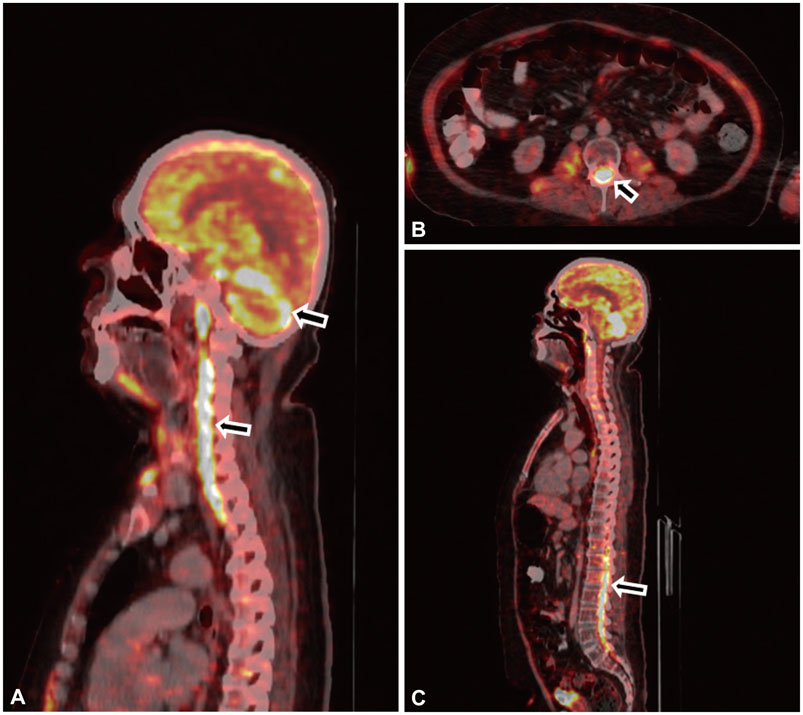
Fig. 4 FDG-PET images (patient 5). A: Intense diffuse FDG uptake (white arrows) in the periphery of both cerebellar hemispheres and the cervical cord. B and C: Intense diffuse FDG uptake (white arrows) in the thickened spinal cord in axial and saggittal sections, respectively (maximum standard uptake value of 25.9) (patient 5). FDG-PET: fluorodeoxyglucose positron tomography.
Cited by 3 articles
-
Partial Conduction Block as an Early Nerve Conduction Finding in Neurolymphomatosis
Hyung Jun Park, Ha Young Shin, Se Hoon Kim, Ha-Neul Jeong, Young-Chul Choi, Bum Chun Suh, Kee Duk Park, Seung Min Kim
J Clin Neurol. 2018;14(1):73-80. doi: 10.3988/jcn.2018.14.1.73.Neurolymphomatosis presenting as brachial plexopathy with involvement of cranial nerves
Hye Jung Lee, Keun Soo Kim, Pamela Song, Jae-Jung Lee, Jung-Joon Sung, Kyomin Choi, Bohyun Kim, Joong-Yang Cho
Ann Clin Neurophysiol. 2018;20(1):44-48. doi: 10.14253/acn.2018.20.1.44.Sciatic nerve neurolymphomatosis as the initial presentation of primary diffuse large B-cell lymphoma: a rare cause of leg weakness
Kyoung Tae Kim, Se Il Kim, Young Rok Do, Hye Ra Jung, Jang Hyuk Cho
Yeungnam Univ J Med. 2021;38(3):258-263. doi: 10.12701/yujm.2021.00983.
Reference
-
1. Petluri G, Goyal MK, Singla V, Mittal BR, Nahar U, Modi M, et al. Neurolymphomatosis: a rare cause of multiple mononeuropathy. World J Neurosci. 2014; 4:190–193.2. Peterson J, Caliskan B, Bonyadlou S. Positron emission tomography/ computerized tomography imaging of multiple focus of neurolymphomatosis. Indian J Nucl Med. 2014; 29:252–253.
Article3. Vallat JM, De Mascarel HA, Bordessoule D, Jauberteau MO, Tabaraud F, Gelot A, et al. Non-Hodgkin malignant lymphomas and peripheral neuropathies--13 cases. Brain. 1995; 118(Pt 5):1233–1245.
Article4. Viala K, Béhin A, Maisonobe T, Léger JM, Stojkovic T, Davi F, et al. Neuropathy in lymphoma: a relationship between the pattern of neuropathy, type of lymphoma and prognosis? J Neurol Neurosurg Psychiatry. 2008; 79:778–782.
Article5. Vital C, Vital A, Julien J, Rivel J, deMascarel A, Vergier B, et al. Peripheral neuropathies and lymphoma without monoclonal gammopathy: a new classification. J Neurol. 1990; 237:177–185.
Article6. Briani C, Vitaliani R, Grisold W, Honnorat J, Graus F, Antoine JC, et al. Spectrum of paraneoplastic disease associated with lymphoma. Neurology. 2011; 76:705–710.
Article7. Baehring JM, Batchelor TT. Diagnosis and management of neurolymphomatosis. Cancer J. 2012; 18:463–468.
Article8. Grisariu S, Avni B, Batchelor TT, van den, Bokstein F, Schiff D, et al. Neurolymphomatosis: an International Primary CNS Lymphoma Collaborative Group report. Blood. 2010; 115:5005–5011.
Article9. Baehring JM, Damek D, Martin EC, Betensky RA, Hochberg FH. Neurolymphomatosis. Neuro Oncol. 2003; 5:104–115.
Article10. Jellinger K, Radiaszkiewicz T. Involvement of the central nervous system in malignant lymphomas. Virchows Arch A Pathol Anat Histol. 1976; 370:345–362.
Article11. Tomita M, Koike H, Kawagashira Y, Iijima M, Adachi H, Taguchi J, et al. Clinicopathological features of neuropathy associated with lymphoma. Brain. 2013; 136(Pt 8):2563–2578.
Article12. Young RC, Howser DM, Anderson T, Fisher RI, Jaffe E, DeVita VT Jr. Central nervous system complications of non-Hodgkin’s lymphoma. The potential role for prophylactic therapy. Am J Med. 1979; 66:435–443.13. Cheson BD. Role of functional imaging in the management of lymphoma. J Clin Oncol. 2011; 29:1844–1854.
Article14. Salm LP, Van der, Stokkel MP. Increasing importance of 18FFDG PET in the diagnosis of neurolymphomatosis. Nucl Med Commun. 2012; 33:907–916.
Article15. Jung YH, Woo IS, Han DJ, Han CW. (18)F-fluoro-2-deoxy-D-glucose positron emission tomography findings of neurolymphomatosis. Blood Res. 2014; 49:83.
Article16. Cheung C, Lopes D, Hung KN, Chan T, Chan KW, Kwong YL. Neurolymphomatosis: role of positron emission tomography in diagnosis. Ann Hematol. 2012; 91:1313–1314.
Article17. Hong CM, Lee SW, Lee HJ, Song BI, Kim HW, Kang S, et al. Neurolymphomatosis on F-18 FDG PET/CT and MRI findings: a case report. Nucl Med Mol Imaging. 2011; 45:76–78.
Article
- Full Text Links
-
- Actions
-
Cited
- CITED
-
- Close
- Share
-
- Similar articles
-
- Neurolymphomatosis Involving Sciatic Nerve: A Case Report
- Neurolymphomatosis in a patient with T-cell non-Hodgkin's lymphoma
- Neurolymphomatosis Involving Antebrachial Cutaneous Nerve
- Lumbosacral plexopathy due to neurolymphomatosis superimposed on traumatic nerve injury
- A Case of Neurolymphomatosis Involving Cranial Nerve Diagnosed by PET-CT Imaging
