The Change of Lacrimal Gland Volume in Korean Patients with Thyroid-associated Ophthalmopathy
- Affiliations
-
- 1Department of Ophthalmology, Gyeongsang National University College of Medicine, Jinju, Korea. stramast@naver.com
- 2Gyeongsang Institute of Health Science, Gyeongsang National University, Jinju, Korea.
- KMID: 2353824
- DOI: http://doi.org/10.3341/kjo.2016.30.5.319
Abstract
- PURPOSE
To describe the change of lacrimal gland volumes in Korean patients with thyroid-associated ophthalmopathy (TAO) via computed tomography (CT).
METHODS
A retrospective review of CT images from 217 TAO patients and 135 control subjects was performed. The TAO patients were diagnosed between May 2005 and May 2014 and had a CT performed on initial presentation (330 orbital CT scans). These images were compared with 270 orbital CT scans from the control group, obtained between May 2013 and May 2014. An open source DICOM viewer was used to calculate the volume of the lacrimal gland.
RESULTS
The mean volume of the lacrimal gland in TAO patients was 0.816 cm³ in the right orbit (standard deviation [SD], 0.048) and 0.811 cm3 in the left orbit (SD, 0.051), with no significant difference between right and left (p = 0.192). However, significant differences were observed between TAO patients and healthy individuals (p < 0.001). There was no significant difference between mean lacrimal gland volumes of males (0.812 cm³; SD, 0.037) and females (0.816 cm³; SD, 0.029) (p = 0.513). There was a negative correlation between gland volume and age in TAO patients (Pearson r = -0.479, p = 0.00). The subjective tearing (right: r = 0.244, p = 0.018; left: r = 0.226, p = 0.024), corneal superficial punctate keratopathy (right: r = 0.192, p = 0.040; left: r = 0.206, p = 0.036), and exophthalmometry (right: r = 0.182, p = 0.032; left: r = 0.180, p = 0.046) correlated with lacrimal gland volume.
CONCLUSIONS
This study is the first to use CT images to calculate the lacrimal gland volume of Korean TAO patients. In TAO patients, the lacrimal gland volume was notably increased compared to control subjects. The lacrimal gland volume decreased with age, but there was no difference between gender and no difference between left and right. The lacrimal gland volume correlated with subjective tearing, corneal superficial punctate keratopathy and exophthalmometry.
Keyword
MeSH Terms
Figure
-
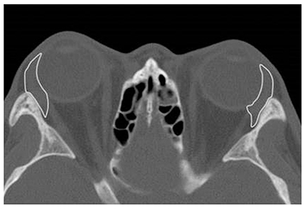
Fig. 1 Axial computed tomography scan viewed on the DICOM viewer (OsiriX, Geneva, Switzerland) with the entire lacrimal gland outlined.

Fig. 2 Scatter plot and correlation between age and average lacrimal gland volume (cm3) in thyroid-associated ophthalmopathy (TAO) patients. B means an inverse relationship between gland volume and age in TAO patients; Pearson r = -0.479 (p = 0.00). A means an inverse relationship between gland volume and age in male TAO patients; Pearson r = -0.328 (p = 0.00). C means an inverse relationship between gland volume and age in female TAO patients; Pearson r = -0.588 (p = 0.00).

Fig. 3 Scatter plot and correlation between age and average lacrimal gland volume (cm3) in control subjects. B means an inverse relationship between gland volume and age in control subjects; Pearson r = -0.802 (p = 0.00). A means an inverse relationship between gland volume and age in male control subjects; Pearson r = -0.772 (p = 0.00). C means an inverse relationship between gland volume and age in female control subjects; Pearson r = -0.830 (p = 0.00).
Reference
-
1. Bahn RS, Dutton CM, Natt N, et al. Thyrotropin receptor expression in Graves' orbital adipose/connective tissues: potential autoantigen in Graves' ophthalmopathy. J Clin Endocrinol Metab. 1998; 83:998–1002.2. Bothun ED, Scheurer RA, Harrison AR, Lee MS. Update on thyroid eye disease and management. Clin Ophthalmol. 2009; 3:543–551.3. Chandler JW, Gillette TE. Immunologic defense mechanisms of the ocular surface. Ophthalmology. 1983; 90:585–591.4. Schaumberg DA, Nichols JJ, Papas EB, et al. The international workshop on meibomian gland dysfunction: report of the subcommittee on the epidemiology of, and associated risk factors for, MGD. Invest Ophthalmol Vis Sci. 2011; 52:1994–2005.5. Matheis N, Grus FH, Breitenfeld M, et al. Proteomics differentiate between thyroid-associated orbitopathy and dry eye syndrome. Invest Ophthalmol Vis Sci. 2015; 56:2649–2656.6. Selter JH, Gire AI, Sikder S. The relationship between Graves' ophthalmopathy and dry eye syndrome. Clin Ophthalmol. 2014; 9:57–62.7. Ismailova DS, Fedorov AA, Grusha YO. Ocular surface changes in thyroid eye disease. Orbit. 2013; 32:87–90.8. Gurdal C, Sarac O, Genc I, et al. Ocular surface and dry eye in Graves' disease. Curr Eye Res. 2011; 36:8–13.9. Gupta A, Sadeghi PB, Akpek EK. Occult thyroid eye disease in patients presenting with dry eye symptoms. Am J Ophthalmol. 2009; 147:919–923.10. An SH, Jin SW, Yang WS, Ahn HB. Calculated brain CT angiography volumes of lacrimal glands in normal Korean orbits. J Korean Ophthalmol Soc. 2014; 55:1413–1417.11. Min SG, Ha MS. Calculated CT volumes of lacrimal glands in normal Korean orbits. J Korean Ophthalmol Soc. 2015; 56:1–5.12. Bingham CM, Harris MA, Realini T, et al. Calculated computed tomography volumes of lacrimal glands and comparison to clinical findings in patients with thyroid eye disease. Ophthal Plast Reconstr Surg. 2014; 30:116–118.13. Bingham CM, Castro A, Realini T, et al. Calculated CT volumes of lacrimal glands in normal Caucasian orbits. Ophthal Plast Reconstr Surg. 2013; 29:157–159.14. Landis JR, Koch GG. The measurement of observer agreement for categorical data. Biometrics. 1977; 33:159–174.15. Kim JH, Lee TS. A study of factors related to the course of graves' ophthalmopathy. J Korean Ophthalmol Soc. 2011; 52:255–260.16. Tamboli DA, Harris MA, Hogg JP, et al. Computed tomography dimensions of the lacrimal gland in normal Caucasian orbits. Ophthal Plast Reconstr Surg. 2011; 27:453–456.17. Harris MA, Realini T, Hogg JP, Sivak-Callcott JA. CT dimensions of the lacrimal gland in Graves orbitopathy. Ophthal Plast Reconstr Surg. 2012; 28:69–72.18. Ueno H, Ariji E, Izumi M, et al. MR imaging of the lacrimal gland: age-related and gender-dependent changes in size and structure. Acta Radiol. 1996; 37:714–719.19. Avetisov SE, Kharlap SI, Markosian AG, et al. Ultrasound spatial clinical analysis of the orbital part of the lacrimal gland in health. Vestn Oftalmol. 2006; 122:14–16.20. Prager A. Macroscopic and microscopic investigations on senile atrophy of the lacrimal gland (preliminary report). Bibl Ophthalmol. 1966; 69:146–158.21. Obata H, Yamamoto S, Horiuchi H, Machinami R. Histopathologic study of human lacrimal gland: statistical analysis with special reference to aging. Ophthalmology. 1995; 102:678–686.22. Gilbard JP, Farris RL. Ocular surface drying and tear film osmolarity in thyroid eye disease. Acta Ophthalmol (Copenh). 1983; 61:108–116.23. Eckstein AK, Finkenrath A, Heiligenhaus A, et al. Dry eye syndrome in thyroid-associated ophthalmopathy: lacrimal expression of TSH receptor suggests involvement of TSHR-specific autoantibodies. Acta Ophthalmol Scand. 2004; 82(3 Pt 1):291–297.24. Crowe JP, Christensen E, Butler J, et al. Primary biliary cirrhosis: the prevalence of hypothyroidism and its relationship to thyroid autoantibodies and sicca syndrome. Gastroenterology. 1980; 78:1437–1441.25. Planck T, Shahida B, Parikh H, et al. Smoking induces overexpression of immediate early genes in active Graves' ophthalmopathy. Thyroid. 2014; 24:1524–1532.26. Wiersinga WM. Smoking and thyroid. Clin Endocrinol (Oxf). 2013; 79:145–151.
- Full Text Links
-
- Actions
-
Cited
- CITED
-
- Close
- Share
-
- Similar articles
-
- A Case of Graves' Disease Showing a Triad of Ophthalmopathy, Pretibial Myxedema and Thyroid Acropachy
- The Natural Course of Strabismus associated with Thyroid Ophthalmopathy
- The Effect of Previous Orbital Decompression on Outcome of Strabismus Surgery in Patients with Thyroid Ophthalmopathy
- Strabismus Surgery for Thyroid Ophthalmopathy
- Calculated CT Volumes of Lacrimal Glands in Normal Korean Orbits
