J Korean Soc Radiol.
2016 Oct;75(4):296-299. 10.3348/jksr.2016.75.4.296.
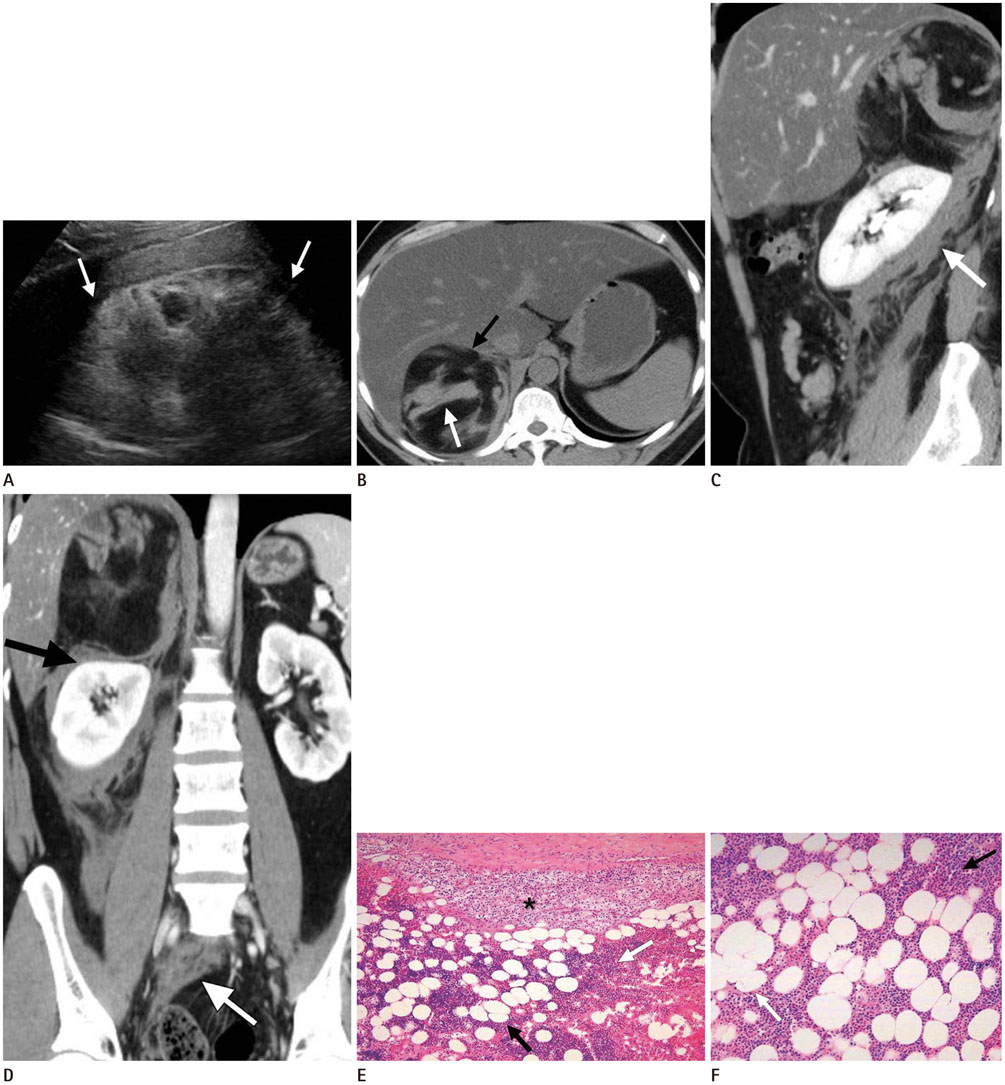
Adrenal Myelolipoma Presenting with Spontaneous Rupture and Retroperitoneal Hemorrhage: A Case Report
- Affiliations
-
- 1Department of Radiology, Eulji Hospital, Eulji University School of Medicine, Seoul, Korea. jyy@eulji.ac.kr
- 2Department of Pathology, Eulji Hospital, Eulji University School of Medicine, Seoul, Korea.
- 3Department of Surgery, Eulji Hospital, Eulji University School of Medicine, Seoul, Korea.
- KMID: 2353680
- DOI: http://doi.org/10.3348/jksr.2016.75.4.296
Abstract
- Adrenal myelolipoma is a rare, benign, and nonfunctioning tumor, which is generally small and asymptomatic and incidentally discovered at autopsy or on radiological examination. In rare cases, large tumors can produce symptoms due to their mass effect or spontaneous rupture with hemorrhage. We reported a case of a 36-year-old man who presented with retroperitoneal hemorrhage due to spontaneous rupture of a large adrenal myelolipoma discovered using multiple imaging modalities.
MeSH Terms
Figure
Reference
-
1. Kenney PJ, Wagner BJ, Rao P, Heffess CS. Myelolipoma: CT and pathologic features. Radiology. 1998; 208:87–95.2. Amano T, Takemae K, Niikura S, Kouno M, Amano M. Retroperitoneal hemorrhage due to spontaneous rupture of adrenal myelolipoma. Int J Urol. 1999; 6:585–588.3. Lattin GE Jr, Sturgill ED, Tujo CA, Marko J, Sanchez-Maldonado KW, Craig WD, et al. From the radiologic pathology archives: Adrenal tumors and tumor-like conditions in the adult: radiologic-pathologic correlation. Radiographics. 2014; 34:805–829.4. Kumar S, Jayant K, Prasad S, Agrawal S, Parma KM, Roat R, et al. Rare adrenal gland emergencies: a case series of giant myelolipoma presenting with massive hemorrhage and abscess. Nephrourol Mon. 2015; 7:e22671.5. Lam KY, Lo CY. Adrenal lipomatous tumours: a 30 year clinicopathological experience at a single institution. J Clin Pathol. 2001; 54:707–712.6. Alvarez JF, Goldstein L, Samreen N, Beegle R, Carter C, Shaw A, et al. Giant adrenal myelolipoma. J Gastrointest Surg. 2014; 18:1716–1718.7. Russell C, Goodacre BW, vanSonnenberg E, Orihuela E. Spontaneous rupture of adrenal myelolipoma: spiral CT appearance. Abdom Imaging. 2000; 25:431–434.8. Gamss C, Chia F, Chernyak V, Rozenblit A. Giant hemorrhagic myelolipoma in a patient with sickle cell disease. Emerg Radiol. 2009; 16:319–322.9. Song JH, Chaudhry FS, Mayo-Smith WW. The incidental adrenal mass on CT: prevalence of adrenal disease in 1,049 consecutive adrenal masses in patients with no known malignancy. AJR Am J Roentgenol. 2008; 190:1163–1168.10. Marti JL, Millet J, Sosa JA, Roman SA, Carling T, Udelsman R. Spontaneous adrenal hemorrhage with associated masses: etiology and management in 6 cases and a review of 133 reported cases. World J Surg. 2012; 36:75–82.
- Full Text Links
- Actions
-
Cited
- CITED
-
- Close
- Share
- Similar articles
-
- Retroperitoneal Hemorrhage due to Spontaneous Rupture of Adrenal Myelolipoma: A case report
- Endovascular Treatment of Massive Retroperitoneal Hemorrhage Due to Spontaneous Rupture of Right Adrenal Gland Metastasis that was Secondary to Invasive Hepatocellular Carcinoma: A Case Report
- Retroperitoneal Hepatocellular Carcinoma Rupture Mimicking an Adrenal Hematoma
- Two Cases of Spontaneous Hemorrhage from Adrenal Tumor
- Surgical management of giant adrenal myelolipoma using a modified Makuuchi incision: a case report


