Cerebrospinal Fluid Pressure and Trans-lamina Cribrosa Pressure Difference in Open-angle Glaucoma: KNHANES V
- Affiliations
-
- 1Department of Ophthalmology, Bucheon Hospital, Soonchunhyang University College of Medicine, Bucheon, Korea. ophkh@schmc.ac.kr
- 2Department of Ophthalmology, Seoul Hospital, Soonchunhyang University College of Medicine, Seoul, Korea.
- KMID: 2351869
- DOI: http://doi.org/10.3341/jkos.2016.57.9.1392
Abstract
- PURPOSE
To investigate the relationships between estimated cerebrospinal fluid pressure (CSFP) and trans-lamina cribrosa pressure difference (TLCPD) in open-angle glaucoma (OAG) in Korean population.
METHODS
A total of 10,801 eyes were included from the Korean National Health and Nutrition Examination Survey V. All participants (aged 19 years or older) were classified as non-glaucomatous group, OAG suspect group and OAG group. CSFP was calculated as CSFP (mm Hg) = 0.44 body mass index (kg/m²) + 0.16 diastolic blood pressure (mm Hg) - 0.18 age (years) - 1.91. TLCPD was calculated by subtracting CSFP from intraocular pressure.
RESULTS
The mean estimated CSFP was (8.7 ± 3.3 mm Hg vs. 11.6 ± 3.7 mm Hg, 11.2 ± 3.8 mm Hg vs. 11.6 ± 3.7 mm Hg) was lower, and the mean TLCPD (5.7 ± 4.4 mm Hg vs. 2.2 ± 4.4 mm Hg, 3 ± 4.7 mm Hg vs. 2.2 ± 4.4 mm Hg) was higher in the OAG group and in the OAG suspect group than in the non-glaucomatous control group, respectively (p < 0.001). After adjusting relating factor with CSFP and TLCPD using simple linear regression and multivariate analyses, the mean estimated CSFP was distributed lower (p < 0.001; beta: -0.12; B: -2.306; 95% confidence interval [CI]: -2.717, -1.895) in OAG group than in non-glaucomatous group and the mean TLCPD was distributed higher (p < 0.001; beta: 0.099; B: 1.349; 95% CI: 0.977, 1.72; p < 0.001; beta: 0.118; B: 2.776; 95% CI: 2.264, 3.289) in OAG suspect group and in OAG group than in non-glaucomatous group, respectively.
CONCLUSIONS
Estimated CSFP and calculated TLCPD showed essential association with OAG presence. It supports the potential role of low CSFP in the pathogenesis of OAG.
Keyword
MeSH Terms
Figure
-
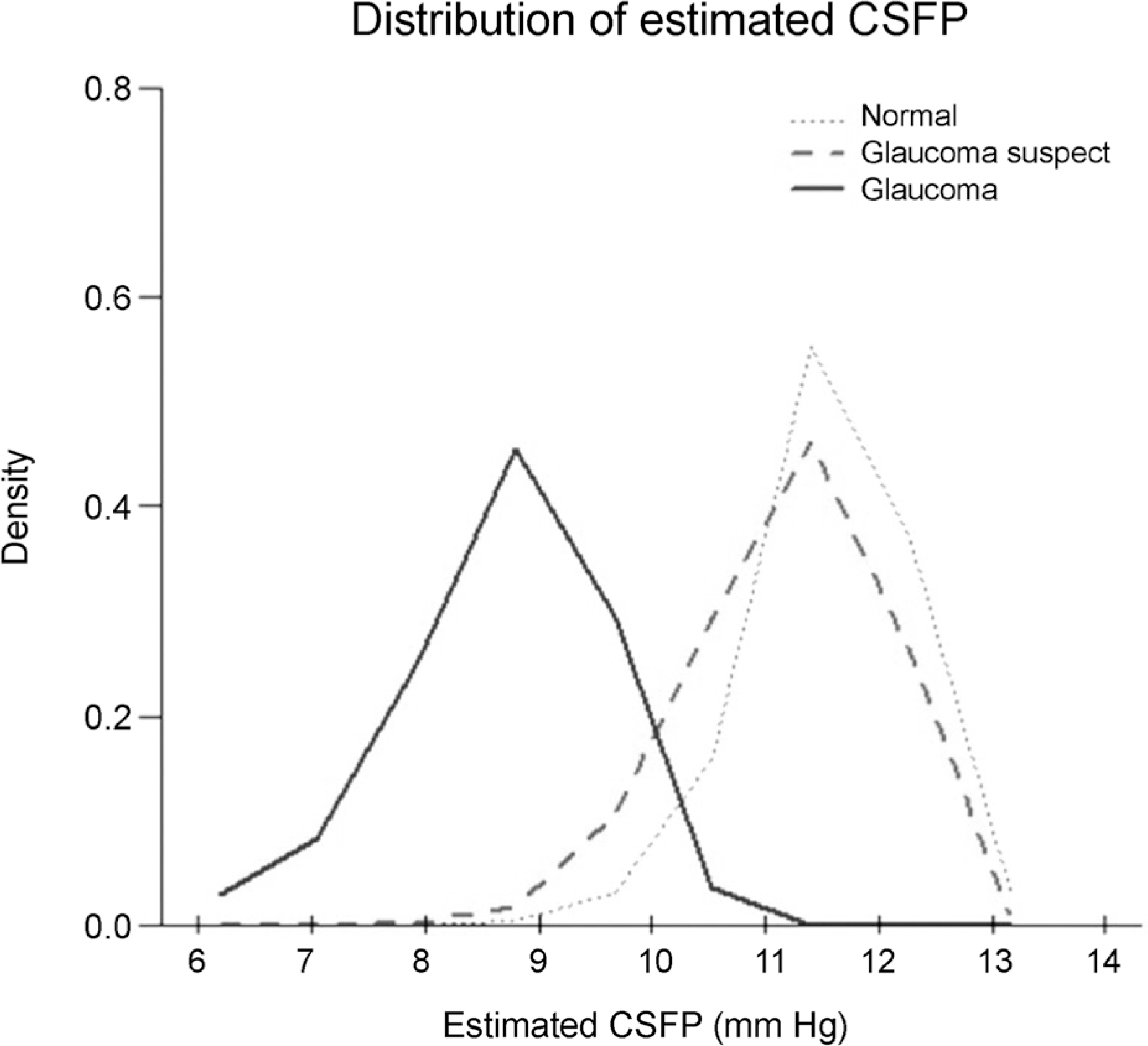
Figure 1. Distribution of estimated cerebrospinal fluid pressure. Estimated cerebrospinal fluid pressure (CSFP) was calculated after adjustment for glaucoma status, sex, log-transformed pulse, total cholesterol, log-transformed triglycerides, corneal refractive power, anterior chamber depth, vertical cup-to-ratio, and intraocular pressure.

Figure 2. Distribution of estimated trans-lamina cribrosa pressure difference. Estimated trans-lamina cribrosa pressure difference (TLCPD) was calculated after adjustment for glaucoma status, sex, log-transformed pulse, corneal refractive power, anterior chamber depth, and vertical cup-to-disc ratio.
Cited by 1 articles
-
Associations between Intraocular Pressure and Systemic Parameters according to the KNHNES 2008-2011
Ji Young Lee, Hye Bin Yim, Kwi Young Kang, Na Young Lee
J Korean Ophthalmol Soc. 2017;58(4):430-436. doi: 10.3341/jkos.2017.58.4.430.
Reference
-
References
1. Crawford Downs J, Roberts MD, Sigal IA. Glaucomatous cupping of the lamina cribrosa: a review of the evidence for active abdominal remodeling as a mechanism. Exp Eye Res. 2011; 93:133–40.2. Yablonski M, Ritch R, Pokorny KS. Effect of decreased abdominal pressure on optic disk. Invest Ophthalmol Vis Sci. 1979; 18(Suppl):165.3. Jonas JB, Berenshtein E, Holbach L. Anatomic relationship abdominal lamina cribrosa, intraocular space, and cerebrospinal fluid space. Invest Ophthalmol Vis Sci. 2003; 44:5189–95.4. Morgan WH, Yu DY, Cooper RL, et al. The influence of abdominal fluid pressure on the lamina cribrosa tissue pressure gradient. Invest Ophthalmol Vis Sci. 1995; 36:1163–72.5. Morgan WH, Chauhan BC, Yu DY, et al. Optic disc movement with variations in intraocular and cerebrospinal fluid pressure. Invest Ophthalmol Vis Sci. 2002; 43:3236–42.6. Morgan WH, Yu DY, Alder VA, et al. The correlation between cerebrospinal fluid pressure and retrolaminar tissue pressure. Invest Ophthalmol Vis Sci. 1998; 39:1419–28.7. Marek B, Harris A, Kanakamedala P, et al. Cerebrospinal fluid pressure and glaucoma: regulation of trans-lamina cribrosa pressure. Br J Ophthalmol. 2014; 98:721–5.
Article8. Ren R, Jonas JB, Tian G, et al. Cerebrospinal fluid pressure in abdominal: a prospective study. Ophthalmology. 2010; 117:259–66.9. Xie X, Zhang X, Fu J, et al. Noninvasive intracranial pressure abdominal by orbital subarachnoid space measurement: the Beijing Intracranial and Intraocular Pressure (iCOP) study. Crit Care. 2013; 17:R162.10. Jonas JB, Nangia V, Wang N, et al. Trans-lamina cribrosa pressure difference and open-angle glaucoma. The central India eye and medical study. PLoS One. 2013; 8:e82284.
Article11. Jonas JB, Wang NL, Wang YX, et al. Estimated trans-lamina cribrosa pressure difference versus intraocular pressure as biomarker for open-angle glaucoma. The Beijing Eye Study 2011. Acta Ophthalmol. 2015; 93:e7–e13.
Article12. Wang YX, Jonas JB, Wang N, et al. Intraocular pressure and estimated cerebrospinal fluid pressure. The Beijing Eye Study 2011. PLoS One. 2014; 9:e104267.
Article13. Yoon KC, Mun GH, Kim SD, et al. Prevalence of eye diseases in South Korea: data from the Korea National Health and Nutrition Examination Survey 2008–2009. Korean J Ophthalmol. 2011; 25:421–33.
Article14. Foster PJ, Buhrmann R, Quigley HA, Johnson GJ. The definition and classification of glaucoma in prevalence surveys. Br J Ophthalmol. 2002; 86:238–42.
Article15. Berdahl JP, Allingham RR, Johnson DH. Cerebrospinal fluid pressure is decreased in primary open-angle glaucoma. Ophthalmology. 2008; 115:763–8.
Article16. Berdahl JP, Fautsch MP, Stinnett SS, Allingham RR. Intracranial pressure in primary open angle glaucoma, normal tension glaucoma, and ocular hypertension: a case-control study. Invest Ophthalmol Vis Sci. 2008; 49:5412–8.
Article17. Guidoboni G, Harris A, Carichino L, et al. Effect of intraocular pressure on the hemodynamics of the central retinal artery: a mathematical model. Math Biosci Eng. 2014; 11:523–46.
Article18. Quigley HA, Broman AT. The number of people with glaucoma worldwide in 2010 and 2020. Br J Ophthalmol. 2006; 90:262–7.
Article19. Kim CS, Seong GJ, Lee NH, et al. Prevalence of primary open-angle glaucoma in central South Korea the Namil study. Ophthalmology. 2011; 118:1024–30.
- Full Text Links
-
- Actions
-
Cited
- CITED
-
- Close
- Share
-
- Similar articles
-
- Correlation between Trans-lamina Cribrosa Pressure Difference and Morphologic Parameters of Optic Disc in Normal Tension Glaucoma Patients
- The Relationships of Intraocular Pressure, Cerebrospinal Fluid Pressure, and Trans-lamina Cribrosa Pressure Differences with Myopia
- Reversal of Optic Disc Cupping in Adults with Advanced Glaucoma
- Glaucoma in Leprosy
- Lamina Cribrosa Thickness in the Fellow Eyes of Patients with Unilateral Retinal Vein Occlusion
