J Korean Assoc Oral Maxillofac Surg.
2016 Apr;42(2):127-130. 10.5125/jkaoms.2016.42.2.127.
Single-tooth dento-osseous osteotomy with a computer-aided design/computer-aided manufacturing surgical guide
- Affiliations
-
- 1Department of Oral and Maxillofacial Surgery, National Health Insurance Service Ilsan Hospital, Goyang, Korea.
- 2Department of Orthodontics, National Health Insurance Service Ilsan Hospital, Goyang, Korea. ilsanortho@gmail.com
- KMID: 2351738
- DOI: http://doi.org/10.5125/jkaoms.2016.42.2.127
Abstract
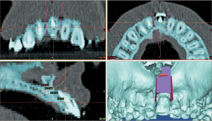
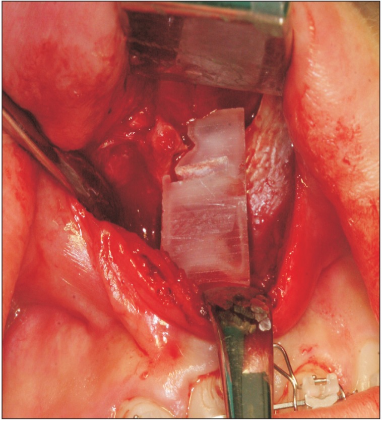
- This clinical note introduces a method to assist surgeons in performing single-tooth dento-osseous osteotomy. For use in this method, a surgical guide was manufactured using computer-aided design/computer-aided manufacturing technology and was based on preoperative surgical simulation data. This method was highly conducive to successful single-tooth dento-osseous segmental osteotomy.
Keyword
MeSH Terms
Figure
Reference
-
1. Rodrigues DB, Wolford LM, Figueiredo LM, Adams GQ. Management of ankylosed maxillary canine with single-tooth osteotomy in conjunction with orthognathic surgery. J Oral Maxillofac Surg. 2014; 72:2419. PMID: 25266594.
Article2. Senşık NE, Koçer G, Kaya BÜ. Ankylosed maxillary incisor with severe root resorption treated with a single-tooth dento-osseous osteotomy, vertical alveolar distraction osteogenesis, and mini-implant anchorage. Am J Orthod Dentofacial Orthop. 2014; 146:371–384. PMID: 25172260.3. Ohkubo K, Susami T, Mori Y, Nagahama K, Takahashi N, Saijo H, et al. Treatment of ankylosed maxillary central incisors by singletooth dento-osseous osteotomy and alveolar bone distraction. Oral Surg Oral Med Oral Pathol Oral Radiol Endod. 2011; 111:561–567. PMID: 21074463.
Article4. Im JJ, Kye MK, Hwang KG, Park CJ. Miniscrew-anchored alveolar distraction for the treatment of the ankylosed maxillary central incisor. Dent Traumatol. 2010; 26:285–288. PMID: 20572845.
Article5. Liem AM, Hoogeveen EJ, Jansma J, Ren Y. Surgically facilitated experimental movement of teeth: systematic review. Br J Oral Maxillofac Surg. 2015; 53:491–506. PMID: 25911054.
Article6. Kang SH, Lee JW, Kim MK. Use of the surface-based registration function of computer-aided design/computer-aided manufacturing software in medical simulation software for three-dimensional simulation of orthognathic surgery. J Korean Assoc Oral Maxillofac Surg. 2013; 39:197–199. PMID: 24471043.
Article
- Full Text Links
- Actions
-
Cited
- CITED
-
- Close
- Share
- Similar articles
-
- Genioplasty using a simple CAD/CAM (computer-aided design and computer-aided manufacturing) surgical guide
- Fabricating a Ceramic-Pressed-to-Metal Restoration with Computer-Aided Design, Computer-Aided Manufacturing and Selective Laser Sintering: A Case Report
- Use of an Extraoral Transfer Jig and a Handheld Face Scanner App for Integrating Face Scan Data into Prosthesis Design
- Recent advances in the reconstruction of cranio-maxillofacial defects using computer-aided design/computer-aided manufacturing
- The application of “bone window technique” using piezoelectric saws and a CAD/CAM-guided surgical stent in endodontic microsurgery on a mandibular molar case


