The association between radiographic embrasure morphology and interdental papilla reconstruction using injectable hyaluronic acid gel
- Affiliations
-
- 1Department of Periodontology, Chosun University School of Dentistry, Gwangju, Korea. bobkim@chosun.ac.kr
- 2Department of Oral and Maxillofacial Radiology, Chosun University School of Dentistry, Gwangju, Korea.
- 3Department of Prosthodontics, Chosun University School of Dentistry, Gwangju, Korea.
- KMID: 2350028
- DOI: http://doi.org/10.5051/jpis.2016.46.4.277
Abstract
- PURPOSE
The purpose of this study was to evaluate the clinical efficacy of enhancing deficient interdental papilla with hyaluronic acid gel injection by assessing the radiographic anatomical factors affecting the reconstruction of the interdental papilla.
METHODS
Fifty-seven treated sites from 13 patients (6 males and 7 females) were included. Patients had papillary deficiency in the upper anterior area. Prior to treatment, photographic and periapical radiographic standardization devices were designed for each patient. A 30-gauge needle was used with an injection-assistance device to inject a hyaluronic acid gel to the involved papilla. This treatment was repeated up to 5 times every 3 weeks. Patients were followed up for 6 months after the initial gel application. Clinical photographic measurements of the black triangle area (BTA), height (BTH), and width (BTW) and periapical radiographic measurements of the contact point and the bone crest (CP-BC) and the interproximal distance between roots (IDR) were undertaken using computer software. The interdental papilla reconstruction rate (IPRR) was calculated to determine the percentage change of BTA between the initial and final examination and the association between radiographic factors and the reconstruction of the interdental papilla by means of injectable hyaluronic acid gel were evaluated.
RESULTS
All sites showed improvement between treatment examinations. Thirty-six sites had complete interdental papilla reconstruction and 21 sites showed improvement ranging from 19% to 96%. The CP-BC correlated with the IPRR. More specifically, when the CP-BC reached 6 mm, virtually complete interdental papilla reconstruction via injectable hyaluronic acid gel was achieved.
CONCLUSIONS
These results suggest that the CP-BC is closely related to the efficacy of hyaluronic acid gel injection for interdental papilla reconstruction.
MeSH Terms
Figure
-

Figure 1 The clinical photographic and periapical radiographic standardization device.
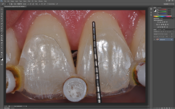
Figure 2 The measurement of clinical photographs using an image analysis system.
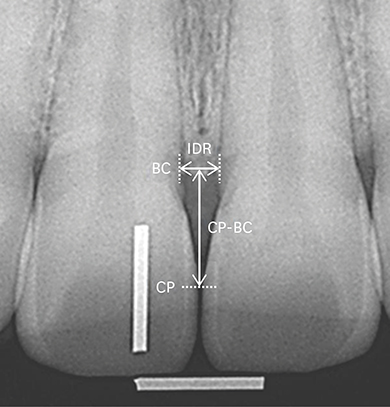
Figure 3 The radiographic analysis. Horizontal and vertical wires of 5-mm length are shown. IDR, the interproximal distance between roots; CP-BC, the distance between the most apical part of the CP (contact point) and the most coronal portion of the BC (bone crest).
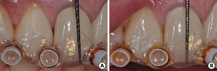
Figure 4 A case of complete interdental papilla reconstruction. (A) At baseline. (B) At 6 months after reconstruction.
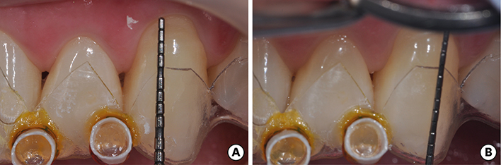
Figure 5 A case of partial interdental papilla reconstruction. (A) At baseline. (B) At 6 months after reconstruction.
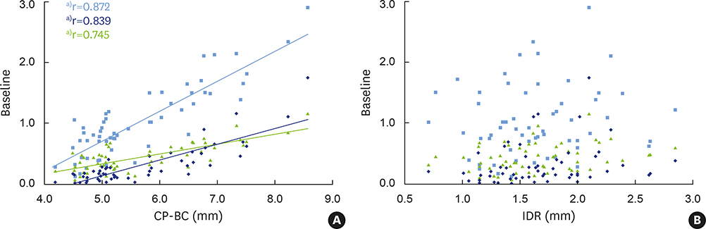
Figure 6 The correlations among the distance between the contact point and the bone crest (CP-BC), the interproximal distance between the roots (IDR), and the baseline black triangle. (A) Statistically significant positive correlation between CP-BC and the baseline black triangle was observed. (B) No correlation was observed between the IDR and the baseline black triangle. , Black triangle area (mm2); , Black triangle height (mm); , Black triangle width (mm). r, Pearson correlation coefficient. a)Statistically significant (P<0.05).
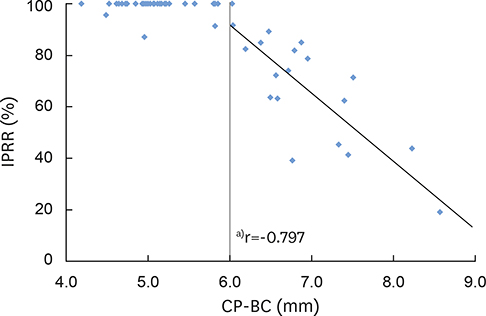
Figure 7 The correlation between the distance between the contact point and the bone crest (CP-BC) and the interdental papilla reconstruction rate (IPRR). Two groups were categorized based on a CP-BC cut-off value of 6 mm. A statistically significant negative correlation of CP-BC>6.0 mm and IPRR was observed. r, Pearson correlation coefficient. a)Statistically significant (P<0.05).
Reference
-
1. Grunder U. The inlay-graft technique to create papillae between implants. J Esthet Dent. 1997; 9:165–168.
Article2. Choquet V, Hermans M, Adriaenssens P, Daelemans P, Tarnow DP, Malevez C. Clinical and radiographic evaluation of the papilla level adjacent to single-tooth dental implants. A retrospective study in the maxillary anterior region. J Periodontol. 2001; 72:1364–1371.
Article3. Tarnow DP, Cho SC, Wallace SS. The effect of inter-implant distance on the height of inter-implant bone crest. J Periodontol. 2000; 71:546–549.
Article4. Tarnow DP, Eskow RN. Considerations for single-unit esthetic implant restorations. Compend Contin Educ Dent. 1995; 16:778–784.5. Tarnow DP, Magner AW, Fletcher P. The effect of the distance from the contact point to the crest of bone on the presence or absence of the interproximal dental papilla. J Periodontol. 1992; 63:995–996.
Article6. Cho HS, Jang HS, Kim DK, Park JC, Kim HJ, Choi SH, et al. The effects of interproximal distance between roots on the existence of interdental papillae according to the distance from the contact point to the alveolar crest. J Periodontol. 2006; 77:1651–1657.
Article7. Chen MC, Liao YF, Chan CP, Ku YC, Pan WL, Tu YK. Factors influencing the presence of interproximal dental papillae between maxillary anterior teeth. J Periodontol. 2010; 81:318–324.
Article8. Kim JH, Cho YJ, Lee JY, Kim SJ, Choi JI. An analysis on the factors responsible for relative position of interproximal papilla in healthy subjects. J Periodontal Implant Sci. 2013; 43:160–167.
Article9. Chang LC. The association between embrasure morphology and central papilla recession. J Clin Periodontol. 2007; 34:432–436.
Article10. Chang LC. Assessment of parameters affecting the presence of the central papilla using a non-invasive radiographic method. J Periodontol. 2008; 79:603–609.
Article11. Beagle JR. Surgical reconstruction of the interdental papilla: case report. Int J Periodontics Restorative Dent. 1992; 12:145–151.12. Jemt T. Regeneration of gingival papillae after single-implant treatment. Int J Periodontics Restorative Dent. 1997; 17:326–333.13. Nemcovsky CE. Interproximal papilla augmentation procedure: a novel surgical approach and clinical evaluation of 10 consecutive procedures. Int J Periodontics Restorative Dent. 2001; 21:553–559.14. Lee EK, Herr Y, Kwon YH, Shin SI, Lee DY, Chung JH. I-shaped incisions for papilla reconstruction in second stage implant surgery. J Periodontal Implant Sci. 2010; 40:139–143.
Article15. Nemcovsky CE, Moses O, Artzi Z. Interproximal papillae reconstruction in maxillary implants. J Periodontol. 2000; 71:308–314.
Article16. Becker W, Gabitov I, Stepanov M, Kois J, Smidt A, Becker BE. Minimally invasive treatment for papillae deficiencies in the esthetic zone: a pilot study. Clin Implant Dent Relat Res. 2010; 12:1–8.
Article17. Necas J, Bartosikova L, Brauner P, Kolar J. Hyaluronic acid (hyaluronan): a review. Vet Med (Praha). 2008; 53:397–411.
Article18. Silness J, Loe H. Periodontal disease in pregnancy. II. Correlation between oral hygiene and periodontal condition. Acta Odontol Scand. 1964; 22:121–135.
Article19. Loe H, Silness J. Periodontal disease in pregnancy. I. Prevalence and severity. Acta Odontol Scand. 1963; 21:533–551.
Article20. Mansouri SS, Ghasemi M, Salmani Z, Shams N. Clinical application of hyaluronic acid gel for reconstruction of interdental papilla at the esthetic zone. J Islam Dent Assoc Iran. 2013; 25:152–157.
- Full Text Links
-
- Actions
-
Cited
- CITED
-
- Close
- Share
-
- Similar articles
-
- Analysis of the embrasure dimensions between maxillary central incisors in relation to the topography of the interdental papilla
- An analysis on the factors responsible for relative position of interproximal papilla in healthy subjects
- Glans Penis Augmentation Using Hyaluronic Acid Gel as an Injectable Filler
- Strategic serial extractions and immediate implantation for interdental papilla preservation: A case report
- Breast Enhancement using Hyaluronic Acid Gel Filler
