Analysis of the embrasure dimensions between maxillary central incisors in relation to the topography of the interdental papilla
- Affiliations
-
- 1Department of Periodontology, Institute of Oral Bioscience, Chonbuk National University School of Dentistry, Jeonju, Korea. chang@chonbuk.ac.kr
- KMID: 2094723
- DOI: http://doi.org/10.5051/jpis.2011.41.6.273
Abstract
- PURPOSE
To analyze the dimensions of the embrasure space between the maxillary central incisors as potential factors influencing interdental papilla fill and height.
METHODS
The embrasure dimensions between the maxillary central incisors of 100 subjects (40 females/60 males) were assessed with clinical, study model, and radiographic examinations. Variables of the complete and deficient papilla fill groups were compared. Multiple regression analyses were performed to investigate potential influence of the distance between the contact point and bone crest (CP_BC), horizontal interdental distance (HID), and facio-lingual thickness (FLT) at the papilla base on complete/deficient papilla fill and papilla height (PH).
RESULTS
CP_BC was the only variable that showed a significant difference between the complete and deficient papilla groups (P<0.05). When the CP_BC was less than 5 mm, the embrasure spaces between the maxillary central incisors were completely filled with interdental papilla. Multiple regression analyses revealed that a significant predictor for complete/deficient papilla fill was CP_BC, and significant predictors for PH were CP_BC and HID (P<0.05).
CONCLUSIONS
The chances of complete papilla fill increased as CP_BC decreased, while PH increased as CP_BC and HID increased. However, the FLT of the papilla base did not appear to affect papilla fill or PH. From an esthetic perspective, CP_BC as well as HID should be considered as factors influencing the topography of interdental papilla.
Keyword
Figure
-
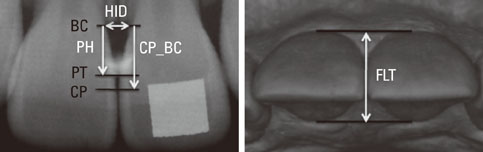
Figure 1 Radiographic and study model assessments of various embrasure dimensions between the maxillary central incisors. HID: horizontal interdental distance, BC: bone crest, PH: papilla height, PT: papilla tip, CP: contact point, CP_BC: distance between the contact point and bone crest, FLT: facio-lingual thickness of the papilla base.
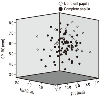
Figure 2 Relationships between complete/deficient papilla and the three embrasure dimensions. CP_BC: distance between the contact point and bone crest, HID: horizontal interdental distance, FLT: facio-lingual thickness at the papilla.

Figure 3 Relationships between papilla height (PH) and 3 embrasure dimensions. (A) Distance between the contact point and bone crest (CP_BC), (B) Horizontal interdental distance (HID), (C) Facio-lingual thickness at the papilla base (FLT). Dotted lines indicate 95% confidence interval for the regression line.
Reference
-
1. Tarnow DP, Magner AW, Fletcher P. The effect of the distance from the contact point to the crest of bone on the presence or absence of the interproximal dental papilla. J Periodontol. 1992. 63:995–996.
Article2. Kurth JR, Kokich VG. Open gingival embrasures after orthodontic treatment in adults: prevalence and etiology. Am J Orthod Dentofacial Orthop. 2001. 120:116–123.
Article3. Cho HS, Jang HS, Kim DK, Park JC, Kim HJ, Choi SH, et al. The effects of interproximal distance between roots on the existence of interdental papillae according to the distance from the contact point to the alveolar crest. J Periodontol. 2006. 77:1651–1657.
Article4. Martegani P, Silvestri M, Mascarello F, Scipioni T, Ghezzi C, Rota C, et al. Morphometric study of the interproximal unit in the esthetic region to correlate anatomic variables affecting the aspect of soft tissue embrasure space. J Periodontol. 2007. 78:2260–2265.
Article5. Chang LC. Assessment of parameters affecting the presence of the central papilla using a non-invasive radiographic method. J Periodontol. 2008. 79:603–609.
Article6. Chen MC, Liao YF, Chan CP, Ku YC, Pan WL, Tu YK. Factors influencing the presence of interproximal dental papillae between maxillary anterior teeth. J Periodontol. 2010. 81:318–324.
Article7. Chow YC, Eber RM, Tsao YP, Shotwell JL, Wang HL. Factors associated with the appearance of gingival papillae. J Clin Periodontol. 2010. 37:719–727.
Article8. Gastaldo JF, Cury PR, Sendyk WR. Effect of the vertical and horizontal distances between adjacent implants and between a tooth and an implant on the incidence of interproximal papilla. J Periodontol. 2004. 75:1242–1246.
Article9. Chang LC. Factors Associated With the Interdental Papilla Height Between Two Maxillary Central Incisors: A Radiographic Study. J Periodontol. 2011. 05. 04. [Epub]. DOI:10.1902/jop.2011.100574.
Article10. Jemt T. Regeneration of gingival papillae after single-implant treatment. Int J Periodontics Restorative Dent. 1997. 17:326–333.11. Ash MM. Ash MM, editor. The Permanent Maxillary Incisors. Wheeler's dental anatomy, physiology and occlusion. 1993. Philadelphia: W.B. Saunders;128–149.12. Teughels W, Merheb J, Quirynen M. Critical horizontal dimensions of interproximal and buccal bone around implants for optimal aesthetic outcomes: a systematic review. Clin Oral Implants Res. 2009. 20:Suppl 4. 134–145.
Article13. Phillips K, Kois JC. Aesthetic peri-implant site development. The restorative connection. Dent Clin North Am. 1998. 42:57–70.14. Wennström JL. Mucogingival considerations in orthodontic treatment. Semin Orthod. 1996. 2:46–54.
Article15. Chang LC. Effect of bone crest to contact point distance on central papilla height using embrasure morphologies. Quintessence Int. 2009. 40:507–513.16. Strebel J, Ender A, Paqué F, Krhäenmann M, Attin T, Schmidlin PR. In vivo validation of a three-dimensional optical method to document volumetric soft tissue changes of the interdental papilla. J Periodontol. 2009. 80:56–61.
Article
- Full Text Links
-
- Actions
-
Cited
- CITED
-
- Close
- Share
-
- Similar articles
-
- Anatomic study of the incisive canal in relation to midpalatal placement of mini-implant
- Strategic serial extractions and immediate implantation for interdental papilla preservation: A case report
- An analysis on the factors responsible for relative position of interproximal papilla in healthy subjects
- Alterations of papilla dimensions after orthodontic closure of the maxillary midline diastema: a retrospective longitudinal study
- A Relationship between Interdental Papilla Existence and the Distance from Contact Point to Interdental Alveolar Crest in the Maxillary Anterior Dentition of Korean adults
