Alterations of papilla dimensions after orthodontic closure of the maxillary midline diastema: a retrospective longitudinal study
- Affiliations
-
- 1Department of Periodontology, Chonbuk National University School of Dentistry and Institute of Oral Bioscience, Jeonju, Korea. chang@chonbuk.ac.kr
- 2Department of Orthodontics, Chonbuk National University School of Dentistry and Institute of Oral Bioscience, Jeonju, Korea.
- 3Research Institute of Clinical Medicine of Chonbuk National University-Biomedical Research Institute of Chonbuk National University Hospital, Jeonju, Korea.
- KMID: 2382983
- DOI: http://doi.org/10.5051/jpis.2016.46.3.197
Abstract
- PURPOSE
The aim of this study was to evaluate alterations of papilla dimensions after orthodontic closure of the diastema between maxillary central incisors.
METHODS
Sixty patients who had a visible diastema between maxillary central incisors that had been closed by orthodontic approximation were selected for this study. Various papilla dimensions were assessed on clinical photographs and study models before the orthodontic treatment and at the follow-up examination after closure of the diastema. Influences of the variables assessed before orthodontic treatment on the alterations of papilla height (PH) and papilla base thickness (PBT) were evaluated by univariate regression analysis. To analyze potential influences of the 3-dimensional papilla dimensions before orthodontic treatment on the alterations of PH and PBT, a multiple regression model was formulated including the 3-dimensional papilla dimensions as predictor variables.
RESULTS
On average, PH decreased by 0.80 mm and PBT increased after orthodontic closure of the diastema (P<0.01). Univariate regression analysis revealed that the PH (P=0.002) and PBT (P=0.047) before orthodontic treatment influenced the alteration of PH. With respect to the alteration of PBT, the diastema width (P=0.045) and PBT (P=0.000) were found to be influential factors. PBT before the orthodontic treatment significantly influenced the alteration of PBT in the multiple regression model.
CONCLUSIONS
PH decreased but PBT increased after orthodontic closure of the diastema. The papilla dimensions before orthodontic treatment influenced the alterations of PH and PBT after closure of the diastema. The PBT increased more when the diastema width before the orthodontic treatment was larger.
MeSH Terms
Figure
-
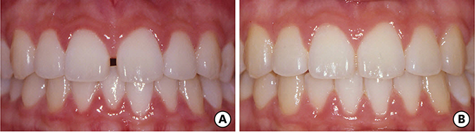
Figure 1 Clinical photographs of a 13-year-old female patient. (A) Before orthodontic closure of the diastema between maxillary central incisors; (B) At the follow-up examination after closure of the diastema.
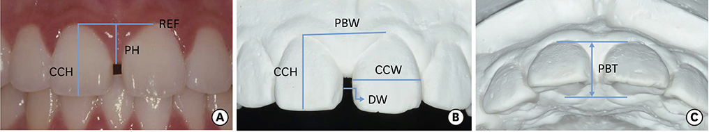
Figure 2 Photographic (A) and study model assessments (B and C). CCH, clinical crown height; PH, papilla height; REF, reference line; PBW, papilla base width; CCW, clinical crown width; DW, diastema width; PBT, papilla base thickness.
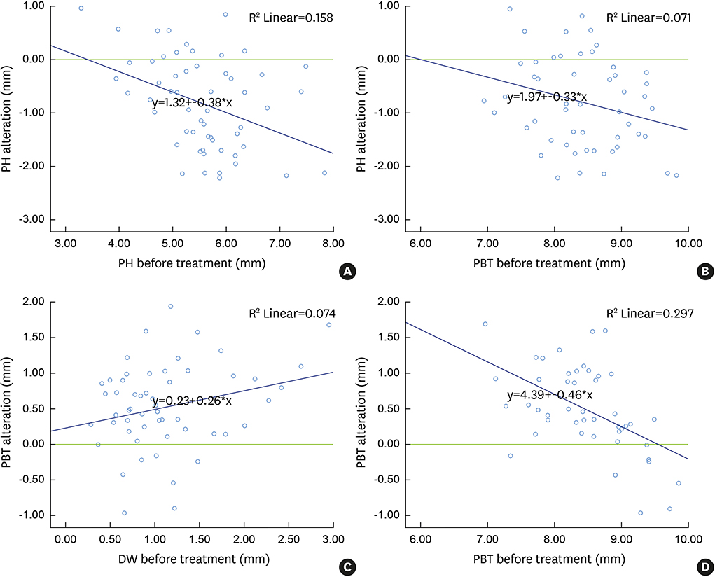
Figure 3 Relationships between the papilla dimensions assessed before orthodontic treatment and the alterations of papilla height and papilla base thickness after orthodontic closure of the diastema. (A) Papilla height versus papilla height alteration; (B) Papilla base thickness versus papilla height alteration; (C) Diastema width versus papilla base thickness alteration; (D) Papilla base thickness versus papilla base thickness alteration. PH, papilla height; PBT, papilla base thickness; DW, diastema width.
Reference
-
1. Chang LC. The association between embrasure morphology and central papilla recession. J Clin Periodontol. 2007; 34:432–436.
Article2. Chang M, Wennström JL. Soft tissue topography and dimensions lateral to single implant-supported restorations. a cross-sectional study. Clin Oral Implants Res. 2013; 24:556–562.
Article3. Kokich VG. Esthetics: the orthodontic-periodontic restorative connection. Semin Orthod. 1996; 2:21–30.
Article4. Prato GP, Rotundo R, Cortellini P, Tinti C, Azzi R. Interdental papilla management: a review and classification of the therapeutic approaches. Int J Periodontics Restorative Dent. 2004; 24:246–255.
Article5. Sharma AA, Park JH. Esthetic considerations in interdental papilla: remediation and regeneration. J Esthet Restor Dent. 2010; 22:18–28.
Article6. Sato S, Nomura N, Kawashima H, Ito K. Creation of a nonsurgical papilla in orthodontic treatment with severe periodontal disease: a case report. Quintessence Int. 2007; 38:e218–e221.7. Kandasamy S, Goonewardene M, Tennant M. Changes in interdental papillae heights following alignment of anterior teeth. Aust Orthod J. 2007; 23:16–23.8. Kim YK, Kwon EY, Cho YJ, Lee JY, Kim SJ, Choi J. Changes in the vertical position of interdental papillae and interseptal bone following the approximation of anterior teeth. Int J Periodontics Restorative Dent. 2014; 34:219–224.
Article9. Cardaropoli G, Lekholm U, Wennström JL. Tissue alterations at implant-supported single-tooth replacements: a 1-year prospective clinical study. Clin Oral Implants Res. 2006; 17:165–171.
Article10. Trentini CM, Moriarty JD, Phillips C, Tulloch JF. Evaluation of the use of orthodontic records to measure the width of keratinized tissue. J Periodontol. 1995; 66:438–442.
Article11. Olsson M, Lindhe J, Marinello CP. On the relationship between crown form and clinical features of the gingiva in adolescents. J Clin Periodontol. 1993; 20:570–577.
Article12. Jemt T. Regeneration of gingival papillae after single-implant treatment. Int J Periodontics Restorative Dent. 1997; 17:326–333.13. Fürhauser R, Florescu D, Benesch T, Haas R, Mailath G, Watzek G. Evaluation of soft tissue around single-tooth implant crowns: the pink esthetic score. Clin Oral Implants Res. 2005; 16:639–644.
Article14. Kokich VO, Kokich VG, Kiyak HA. Perceptions of dental professionals and laypersons to altered dental esthetics: asymmetric and symmetric situations. Am J Orthod Dentofacial Orthop. 2006; 130:141–151.
Article15. Gastaldo JF, Cury PR, Sendyk WR. Effect of the vertical and horizontal distances between adjacent implants and between a tooth and an implant on the incidence of interproximal papilla. J Periodontol. 2004; 75:1242–1246.
Article16. Chow YC, Wang HL. Factors and techniques influencing peri-implant papillae. Implant Dent. 2010; 19:208–219.
Article17. Wennström JL. Mucogingival considerations in orthodontic treatment. Semin Orthod. 1996; 2:46–54.
Article18. Kan JY, Rungcharassaeng K, Umezu K, Kois JC. Dimensions of peri-implant mucosa: an evaluation of maxillary anterior single implants in humans. J Periodontol. 2003; 74:557–562.
Article19. Teughels W, Merheb J, Quirynen M. Critical horizontal dimensions of interproximal and buccal bone around implants for optimal aesthetic outcomes: a systematic review. Clin Oral Implants Res. 2009; 20:Suppl 4. 134–145.
Article
- Full Text Links
-
- Actions
-
Cited
- CITED
-
- Close
- Share
-
- Similar articles
-
- Clinical study on the relapse of diastema
- Diastema closure using direct bonding restorations combined with orthodontic treatment: a case report
- Alterations of the soft tissue dimensions around implant-supported single-tooth replacements in the maxillary anterior region: A retrospective longitudinal study
- Conservative and esthetic closure of maxillary midline diastema without creating "black triangle" using direct resin composite
- Analysis of the embrasure dimensions between maxillary central incisors in relation to the topography of the interdental papilla
