Surgical Planning by 3D Printing for Primary Cardiac Schwannoma Resection
- Affiliations
-
- 1Department of Thoracic and Cardiovascular Surgery, Gachon University Gil Medical Center, Gachon University, Incheon, Korea. lji@gilhospital.com, junyb@gilhospital.com
- 2Department of Molecular Medicine, Graduate School of Medicine, Gachon University, Incheon, Korea.
- KMID: 2345907
- DOI: http://doi.org/10.3349/ymj.2015.56.6.1735
Abstract
- We report herein a case of benign cardiac schwannoma in the interatrial septum. A 42-year-old woman was transferred from a clinic because of cardiomegaly as determined by chest X-ray. A transthoracic echocardiography and chest computed tomography examination revealed a huge mass in the pericardium compressing the right atrium, superior vena cava (SVC), left atrium, and superior pulmonary vein. To confirm that the tumor originated from either heart or mediastinum, cine magnetic resonance imaging was performed, but the result was not conclusive. To facilitate surgical planning, we used 3D printing. Using a printed heart model, we decided that tumor resection under cardiopulmonary bypass (CPB) through sternotomy would be technically feasible. At surgery, a huge tumor in the interatrial septum was confirmed. By incision on the atrial roof between the aorta and SVC, tumor enucleation was performed successfully under CPB. Pathology revealed benign schwannoma. The patient was discharged without complication. 3D printing of the heart and tumor was found to be helpful when deciding optimal surgical approach.
Keyword
MeSH Terms
-
Adult
Atrial Septum/pathology/surgery
Cardiomegaly/*etiology/radiography
*Cardiopulmonary Bypass
Female
Heart Atria/pathology
Heart Neoplasms/pathology/*surgery
Humans
Magnetic Resonance Imaging, Cine
Neurilemmoma/*pathology/surgery
*Printing, Three-Dimensional
Sternotomy
Treatment Outcome
Vena Cava, Superior/pathology
Figure
-
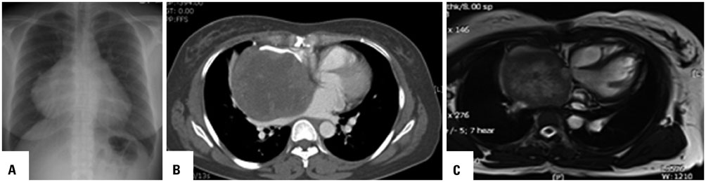
Fig. 1 Chest X-ray and computed tomograph obtained before stent grafting. (A) Chest X-ray showing an extended right heart silhouette. (B) CT revealing a 10×9.5 cm sized heterogeneously enhanced mass in the right pericardial space compressing the superior vena cava, right atrium, left atrium, and superior pulmonary vein. (C) MRI showing a large mass with central necrosis in the right paracardiac area extending to the right side of the ascending aorta between the SVC and right superior PV. SVC, superior vena cava; PV, pulmonary vein.
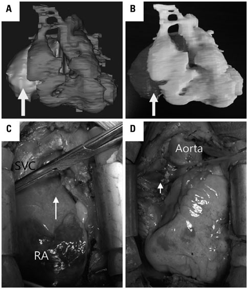
Fig. 2 3D printing model and operating finding. (A) The segmented 3D image using Mimics Base version 16 (Materialize, Leuven, Belgium) showed a huge cardiac mass (arrow). (B) The model of heart and mass which was printed by 3D printer (uPrint, Stratasys Ltd., MN, USA) showed a huge mass (arrow). (C) After sternotomy, a huge yellowish mass was confirmed at Waterstone's groove (arrow). (D) After tumor removal, repair of the interatrial septum was unnecessary, because the tumor did not infiltrate the right or left atrial cavities. Arrow indicates the position where the tumor occupied. SVC, superior vena cava; RA, right atrium.
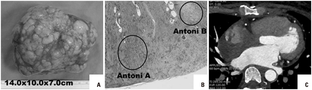
Fig. 3 Pathologic findings and postoperative CT. (A) Macroscopically, the mass was capsulated, nobular, and yellowish, and contained hemorrhagic part. (B) The microscopic finding showed Antoni A area, with hypercellularity composed of palisading nuclei and fibers, and Antoni B area with hypocellularity. (C) Postoperative coronary artery CT showed that there was no remnant tumor.
Reference
-
1. Hwang SK, Jung SH. Schwannoma of the heart. Korean J Thorac Cardiovasc Surg. 2014; 47:141–144.
Article2. Nakamura K, Onitsuka T, Yano M, Yano Y. Surgical resection of right atrial neurilemoma extending to pulmonary vein. Eur J Cardiothorac Surg. 2003; 24:840–842.
Article3. Monroe B, Federman M, Balogh K. Cardiac neurilemoma. Report of a case with electron microscopic examination. Arch Pathol Lab Med. 1984; 108:300–304.4. Jacobs S, Grunert R, Mohr FW, Falk V. 3D-Imaging of cardiac structures using 3D heart models for planning in heart surgery: a preliminary study. Interact Cardiovasc Thorac Surg. 2008; 7:6–9.
Article5. Schmauss D, Haeberle S, Hagl C, Sodian R. Three-dimensional printing in cardiac surgery and interventional cardiology: a single-centre experience. Eur J Cardiothorac Surg. 2015; 47:1044–1052.
Article
- Full Text Links
-
- Actions
-
Cited
- CITED
-
- Close
- Share
-
- Similar articles
-
- Three-Dimensional Printing Technology in Orthopedic Surgery
- Applications of Three-Dimensional Printing in Cardiovascular Surgery: A Case-Based Review
- Clinical Applications of Three-Dimensional Printing in Cardiovascular Disease
- A Review of Current Clinical Applications of Three-Dimensional Printing in Spine Surgery
- Medical Applications of 3D Printing and Standardization Issues
