The Effect of RGP Lens and Reverse Geometry Lens on Redistribution of Corneal Epithelial Cell in Rabbit
- Affiliations
-
- 1Department of Ophthalmology, Kangdong Sacred Heart Hospital, Hallym University College of Medicine, Seoul, Korea. sungpyo@hanafos.com
Abstract
- PURPOSE
To investigate the migration and redistribution of rabbit corneal epithelial cells when wearing reverse geometry lens (RGL) or rigid gas permeable lens (RGP).
METHODS
In 30 rabbits, the right eyes were fitted with either RGL or RGP and the left eyes were untreated to serve as controls. The rabbits were sacrificed at 1, 3, 7, 10 and 14 days after lens fitting. The central and peripheral corneal thicknesses were measured by microscope and the ratio of right to left corneal thickness was calculated to evaluate the characteristics of change over time. By using the molecular probe 7-nitrobenz-2-ox-1,3-diazolylphallacidin (NBD phallacidin), the samples were examined with light microscope to determine the migration and redistribution of epithelial cells in the rabbit cornea.
RESULTS
No consistent changes in the thickness of both central and peripheral corneal epithelium were found. The corneal epithelial cells of both eyes with RGL and RGP reacted positively to NBD phallacidin. The fluorescence was most increased at day 3 of sacrifice in RGL cases and at day 7 in RGP cases, and then decreased in both cases. The corneal epithelium of eyes with RGL exhibited marked increase in the intensity of fluorescence compared to the eyes with RGP.
CONCLUSIONS
The corneal epithelium with RGL showed the strongest intensity of NBD phallacidin fluorescence. This result suggests that wearing RGL may induce the migration and redistribution of corneal epithelial cells.
Keyword
MeSH Terms
Figure
-
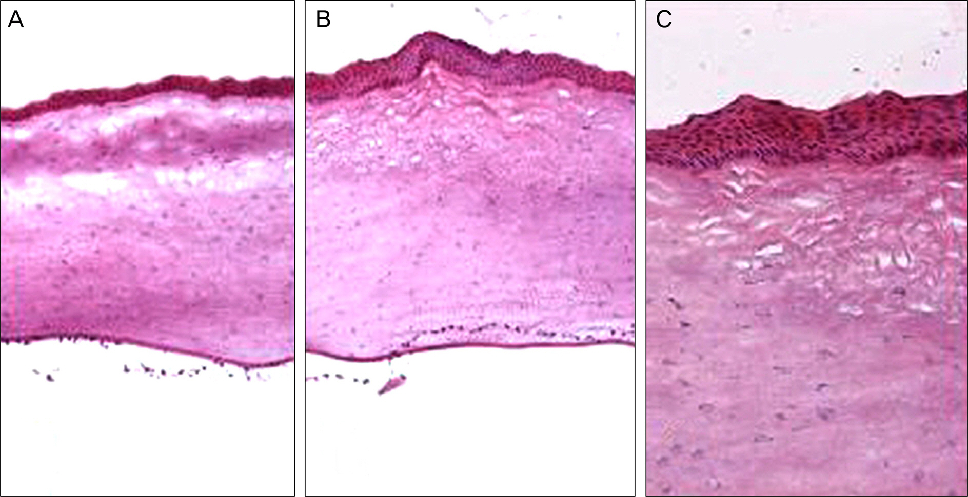
Figure 1. Hematoxylin and eosin (H&E) staining of the central cornea (A) and peripheral cornea (B, C) in reverse geometry lens-wearing rabbit at 10 days (A, B: ×100; C: ×200).
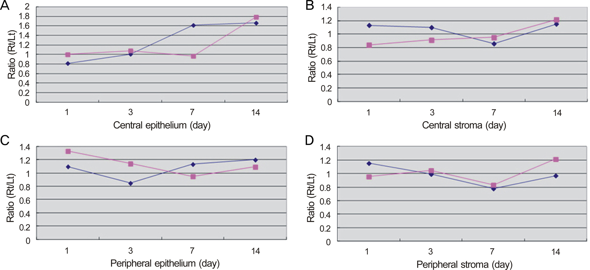
Figure 2. Thickness ratio of each measurement to control in RGL group and RGP group. (A) Central epithelium. (B) Central stroma. (C) Peripheral epithelium. (D) Peripheral stroma.

Figure 3. A positive NBD phallacidin fluorescence of the corneal epithelial cells in each group according to the day of sacrifice after lens fitting. The rabbit in group A was fitted with reverse geometry lens (RGL) and the rabbit in group B was fitted with rigid gas permeable lens (RGP). The photographs show marked increase of NBD phallacidin fluorescence in the corneal epithelial cells in the eyes fitted with RGL. The most increased fluorescence is observed in eyes with RGL at day 3 (arrows). A, B = group; Number = the day of sacrifice.
Reference
-
References
1. Swarbrick HA.Orthokeratology review and update. Clin Exp Optom. 2006; 89:124–43.
Article2. Hiraoka T, Okamoto F, Kaji Y, Oshika T.Optical quality of the cor-nea after overnight orthokeratology. Cornea. 2006; 25(10 Suppl 1):S59–63.
Article3. Stillitano I, Schor P, Lipener C, Hofling-Lima AL.Long-term follow-up of orthokeratology corneal reshaping using wavefront aberrometry and contrast sensitivity. Eye Contact Lens. 2008; 34:140–5.
Article4. Villa-Collar C, González-Méijome JM, Queirós A, Jorge J. Short-term corneal response to corneal refractive therapy for different re-fractive targets. Cornea. 2009; 28:311–6.
Article5. Barr JT, Rah MJ, Jackson JM, Jones LA.Orthokeratology and cor-neal refractive therapy: a review and recent findings. Eye Contact Lens. 2003; 29(1 Suppl):S49–53.
Article6. Hiraoka T, Okamoto C, Ishii Y. . Time course of changes in oc-ular higher-order aberrations and contrast sensitivity after over-night orthokeratology. Invest Ophthalmol Vis Sci. 2008; 49:4314–20.
Article7. Swarbrick HA, Wong G, O'leary DJ. Corneal response to orth- okeratology. Optom Vis Sci. 1998; 75:791–9.8. Nichols JJ, Marsich MM, Nguyen M. . Overnight orth- okeratology. Optom Vis Sci. 2000; 77:252–9.9. Nieto-Bona A, González-Mesa A, Nieto-Bona MP. . Short- term effects of overnight orthokeratology on corneal cell morphol-ogy and corneal thickness. Cornea. 2011; 30:646–54.10. Shin DB, Kim JC, Kim MK. . The effect of RGP lens and re-verse geometry lens on apoptosis in rabbit cornea. J Korean Ophthalmol Soc. 2003; 44:1649–61.11. Shin DB, Lee YW, Kim MK. . The effect of RGP lens and re-verse geometry lens on corneal epithelial proliferation rate in rabbit. J Korean Ophthalmol Soc. 2004; 45:655–67.12. Zhong X, Chen X, Xie RZ. . Differences between overnight and long-term wear of orthokeratology contact lenses in corneal contour, thickness, and cell density. Cornea. 2009; 28:271–9.
Article13. Shin DB, Yang KM, Lee SB. . Effect of reverse geometry lens on correction of moderate-degree myopia and cornea. J Korean Ophthalmol Soc. 2003; 44:1748–56.14. Pollard TD, Aebi U, Cooper JA. . Actin structure, polymer-ization, and gelation. Cold Spring Harb Symp Quant Biol. 1982; 46(Pt 2):513–24.
Article15. Pollard TD.Cytoskeletal functions of cytoplasmic contractile proteins. J Supramol Struct. 1976; 5:317–34.
Article16. Gipson IK, Westcott MJ, Brooksby NG.Effects of cytochalasins B and D and colchicine on migration of the corneal epithelium. Invest Ophthalmol Vis Sci. 1982; 22:633–42.17. Anderson RA.Actin filaments in normal and migrating corneal ep-ithelial cells. Invest Ophthalmol Vis Sci. 1977; 16:161–6.18. Barak LS, Yocum RR, Nothnagel EA, Webb WW.Fluorescence staining of the actin cytoskeleton in living cells with 7-nitro-benz-2-oxa-1,3-diazole-phallacidin. Proc Natl Acad Sci Usa. 1980; 77:980–4.
Article19. Jester JV, Rodrigues MM.Actin filament localization in normal and migrating rabbit corneal epithelium. Curr Eye Res. 1984; 3:955–60.
Article20. Chang JW, Choi TH, Lee HB.The efficacy and safety of reverse geometry lenses. J Korean Ophthalmol Soc. 2004; 45:908–12.21. Soni PS, Nguyen TT, Bonanno JA.Overnight orthokeratology: visual and corneal changes. Eye Contact Lens. 2003; 29:137–45.22. Jester JV, Rodrigues MM, Sun TT.Change in epithelial keratin ex-pression during healing of rabbit corneal wounds. Invest Ophthalmol Vis Sci. 1985; 26:828–37.23. Feng Y, Varikooty J, Simpson TL.Diurnal variation of corneal and corneal epithelial thickness measured using optical coherence tomography. Cornea. 2001; 20:480–3.
Article24. Wang J, Fonn D, Simpson TL, Jones L.Relation between optical coherence tomography and optical pachymetry measurements of corneal swelling induced by hypoxia. Am J Ophthalmol. 2002; 134:93–8.
Article
- Full Text Links
-
- Actions
-
Cited
- CITED
-
- Close
- Share
-
- Similar articles
-
- The Effect of RGP Lens and Reverse Geometry Lens on Apoptosis in Rabbit Cornea
- The Effect of RGP Lens and Reverse Geometry Lens on Corneal Epithelial Proliferation Rate in Rabbit
- How to Improve Success Rate of Rigid Gas Permeable Lens Prescription
- Effect of Reverse Geometry Lens on Correction of Moderate-degree Myopia and Cornea
- Physiological Changes in the Cornea When Wearing Rigid Gas Permeable Contact Lenses
