J Korean Ophthalmol Soc.
2007 Dec;48(12):1630-1635.
Reproducibility of IntraLASIK Flap Thickness Measured with Optical Coherence Tomography
- Affiliations
-
- 1Department of Ophthalmology, Ilsan Paik Hospital, Inje University College of Medicine, Gyeonggi, Korea. jhk0924@hanmail.net
- 2Rhee Eye Hospital, Daejeon, Korea.
Abstract
-
PURPOSE: To investigate the relationship among optical coherence tomography (OCT), ultrasound pachymetry, and Orbscan in central corneal thickness measurement and to evaluate the reproducibility of flap thickness using an IntraLase femtosecond laser.
METHODS
Central corneal thickness was measured by OCT, ultrasound pachymetry, and Orbscan in 59 eyes of 30 patients before LASIK. After IntraLASIK, the corneal flap thickness measured using OCT was compared with the intended corneal flap thickness.
RESULTS
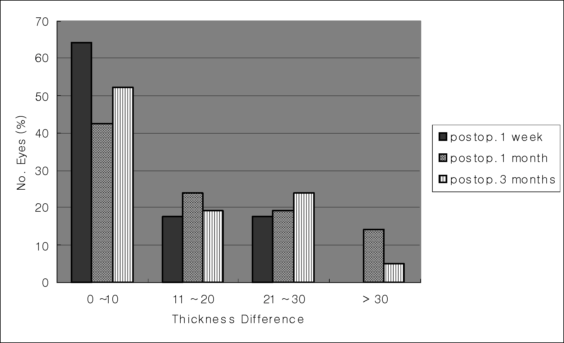
Central corneal thickness measured by OCT was thinner than that measured by other instruments preoperatively, but there was no significant difference among these methods (p>0.01), and corneal thickness values obtained by ultrasound pachymetry and Orbscan correlated well with those obtained by OCT (r ranged from 0.804 to 0.889, p<0.01). After IntraLASIK, there was no significant difference between the mean measured flap thickness and the intended flap thickness (p>0.01).
CONCLUSIONS
OCT is a relatively accurate instrument for measuring corneal thickness and can easily measure the corneal flap thickness after LASIK. Compared with the results of a previous study, the mean measured flap thickness in this study was more reproducible with the IntraLase femtosecond laser.
MeSH Terms
Figure
Reference
-
References
1. Cho HS, Tchah HW. Incidence of Flap Complications of Hansatome (R) in Laser in Situ Keratomileusis. J Korean Ophthalmol Soc. 2003; 43:692–8.2. Seiler T, Koufala K, Richter G. Iatrogenic keratectasia after laser in situ keratmileusis. J Refract Surg. 1998; 14:312–7.3. Arbelaez MC. Nidek MK 2000 microkeratome clinical evaluation. J Refract Surg. 2002; 18:357–60.
Article4. Ucakhan O. Corneal flap thickness in laser in situ keratomileusis using the Summit Krumeich-Barraquer microkeratome. J Cataract Refract Surg. 2002; 28:798–804.5. Shemesh G, Dotan G, Lipshitz I. Predictability of corneal flap thickness in laser in situ keratomileusis using three different microkeratome. J Refract Surg. 2002; 18:347–51.
Article6. Cho WH, Lee DC, Chang MH. Preoperative Factors related to Corneal Flap Thickness in LASIK using Microkeratome. J Korean Ophthalmol Soc. 2006; 47:607–12.7. Jacobs BJ, Deutsch TA, Rubenstein JB. Reproducibility of corneal flap thickness in LASIK. Ophthalmic Surg Lasers. 1999; 30:350–3.
Article8. Yildirim R, Aras C, Ozdamar A, et al. Reproducibility of corneal flap thickness in laser in situ keratomileusis using the Hansatome microkeratome. J Cataract Refract Surg. 2000; 26:1729–32.
Article9. Binder PS. One thounsand consecutive IntraLase laser in situ keratomileusis flaps. J Cataract Refract Surg. 2006; 32:962–9.10. Flanagan GF, Binder PS. Precision of flap measurements for laser in situ keratomileusis in 4428 eyes. J Refract Surg. 2003; 19:113–23.
Article11. Thompson RW Jr, Choi DM, Price MO, et al. Noncontact optical coherence tomography for measurement of corneal flap and residual stromal bed thickness after laser in situ keratomileusis. J Refract Surg. 2003; 19:507–15.
Article12. Eisner RA, Binder PS. Technique for measuring laser in situ keratomileusis flap thickness using the IntraLase laser. J Cataract Refract Surg. 2006; 32:556–8.
Article13. Tomas J, Wang J, Rollins AM, et al. Comparison of corneal thickness measured with Optical Coherence Tomography, Ultrasound pachymetry, and a Scanning slip method. J Refract Surg. 2006; 22:671–8.14. Wang J, Tomas J, Cox I, et al. Noncontact measurements of central corneal epithelial and flap thickness after laser in situ keratomileusis. Invest Ophthalmol Vis Sci. 2004; 45:1812–6.
Article15. Izatt JA, Hee MR, Swanson FA, et al. Micrometer-scale resolution imaging of the anterior eye in vivo optical coherence tomography. Arch Ophthalmol. 1994; 112:1584–9.16. Radhakrishnan S, Rollins AM, Roth JE, et al. Real-time optical coherence tomography of the anterior segment at 1310nm. Arch Ophthalmol. 2001; 119:1179–85.17. Talamo JH, Meltzer J, Gardner J. Reproducibility of flap thickness with IntraLase FS and Moria LSK-1 and M2 microkeratome. J Refract Surg. 2006; 22:556–61.18. Binder PS. Flap dimensions created with the IntraLase FS laser. J Cataract Refract Surg. 2004; 30:26–32.
Article19. Kezirian GM, Stonecipher KG. Comparison of the IntraLase femtosecond laser and mechanical keratomes for laser in situ keratomileusis. J Cataract Refract Surg. 2004; 30:804–11.
Article20. Stahl JE, Durrie DS, Schwendeman FJ, et al. Anterior Segment OCT Aanlysis of Thin IntraLase Femtosecond Flaps. J Refract Surg. 2007; 23:555–8.
- Full Text Links
- Actions
-
Cited
- CITED
-
- Close
- Share
- Similar articles
-
- Comparison of Flap Thickness Measured with Ultrasound Subtraction Method, Direct Method, and Optical Coherence Tomography
- Reproducibility of Choroidal Thickness in Normal Korean Eyes Using Two Spectral Domain Optical Coherence Tomography
- Measurement of Deep Optic Nerve Complex Structures with Two Spectral Domain Optical Coherence Tomography Instruments
- Comparison of Laser In Situ Keratomileusis Flap Morphology and Predictability by WaveLight FS200 Femtosecond Laser and Moria Microkeratome: An Anterior Segment Optical Coherence Tomography Study
- Choroidal Thickness at the Outside of Fovea in Diabetic Retinopathy Using Spectral-Domain Optical Coherence Tomography


