A Study of Joint Space Narrowing and Erosion in Rheumatoid Arthritis
- Affiliations
-
- 1Biomedical Engineering Branch, Division of Cancer, National Cancer Center, Korea. kimkg@ncc.re.kr
- 2Department of Radiology, Yonsei University College of Medicine, Gangnam Severance Hospital, Korea.
Abstract
OBJECTIVE
This study was conducted to measure radiographic joint space width and to estimate erosion in the hands of patients with rheumatoid arthritis. It showed that joint space width, homogeneity, and invariant moments are parameters to discriminate between the normal and the rheumatoid joint. METHODS: In order to measure the joint space width and to estimate erosion in the finger joint, 32 radiographic images were used - 16 images for training and 16 images for testing. The joint space width was measured in order to quantify the joint space narrowing. Also, homogeneity and invariant moments was computed in order to quantify erosion. Finally, artificial neural networks were constructed and tested as a classifier distinguishing between the normal and the rheumatoid joint. RESULTS: The joint space width of normal was 1.04+/-0.15 mm and the width of patients with rheumatoid arthritis was 0.94+/-0.15 mm. The Homogeneity of normal was 16568.83+/-2669.83 and invariant moments were 6843.45+/-2937.55. They were statistically difference (p<.05). Using these characteristics, artificial neural networks showed that they discriminate between normal and rheumatoid arthritis (AUC=0.91). CONCLUSION: Measuring joint space width, estimating homogeneity, and invariant moments provide the capability to distinguish between a normal joint and a rheumatoid joint.
Keyword
Figure
-
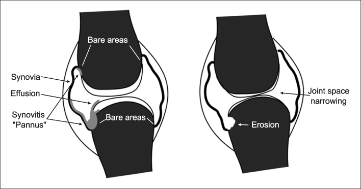
Figure 1 Joint space narrowing and erosive destructions11). An early stage (left), rheumatoid arthritis progression (right)

Figure 2 (A) Original image. (B) The result of median filter
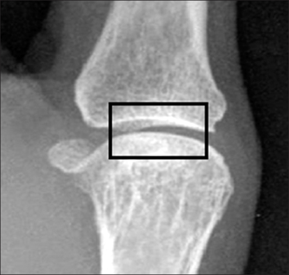
Figure 3 An articulation ROI image

Figure 4 (A) The 2-D LoG function. (B) Discrete approximation to LoG function (σ=1.4)
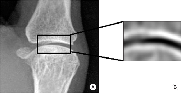
Figure 5 (A) The ROI input image. (B) The result of LoG filter in ROI
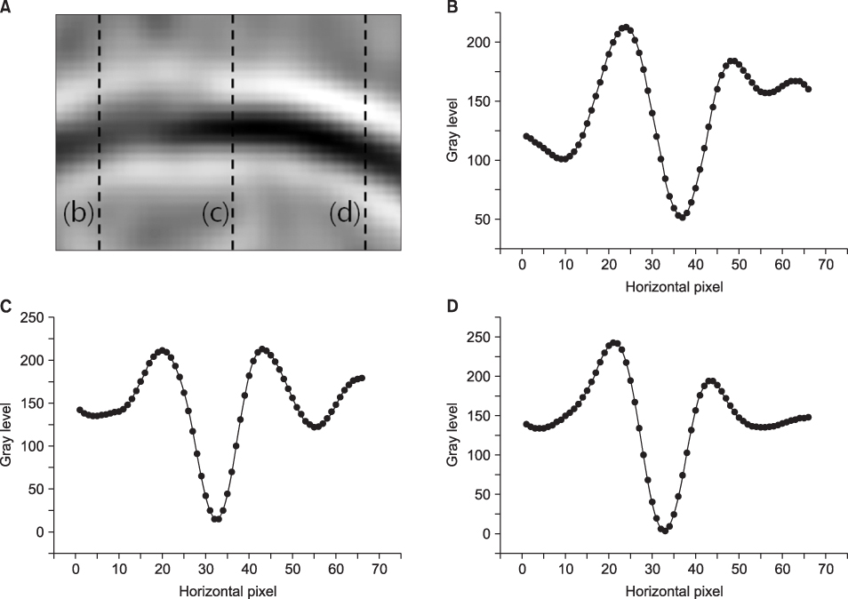
Figure 6 The profiles of ROI. (A) The dot lines are vertical profiles. (B) The profile of the first dot line. (C) The profile of the second dot line. (D) The profile of the third dot line

Figure 7 The distance of a profile

Figure 8 The example of measuring joint space narrowing. (A) The proposed system. (B) The LoG image of the input image. (C) The result image

Figure 9 The box charts of joint space mean width (mm)

Figure 10 (A) Skewness (B) Kurtosis (C) Homogeneity (D) Sum of invariant moments
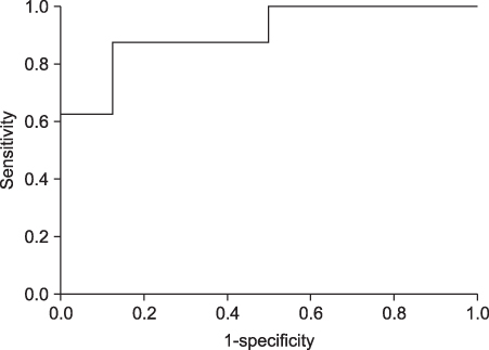
Figure 11 The ROC Curve of ANNs with spatial distance, homogeneity and invariant moments
Reference
-
1. Farrant J, Grainger A, O'Connor P. Advanced imaging in rheumatoid arthritis: part 1: synovitis. Skeletal Radiology. 2007. 36(4):269–279.2. Farrant J, Grainger A, O'Connor P. Advanced imaging in rheumatoid arthritis: part 2: erosion. Skeletal Radiology. 2007. 36(5):381–389.3. Duryea J, Jiang Y, Zakharevich M. Neural network based algorithm to quantify joint space width in joints of the hand for arthritis assessment. Medical Physics. 2000. 27(5):1185–1194.
Article4. Sharp J, Gardner J, Bennett E. Computer-based methods for measuring joint space and estimating erosion volume in the finger and wrist joints of patients with rheumatoid arthritis. Arthritis & Rheumatism. 2000. 43(6):1378–1386.
Article5. Finckh A, Pablo P, Katz J. Performance of an automated computer-based scoring method to assess joint space narrowing in rheumatoid arthritis: a longitudinal study. Arthritis & Rheumatism. 2006. 54(5):1444–1450.
Article6. Sharp J, Bluhm G, Brook Al. Reproducibility of multiple-observer scoring of radiologic abnormalities in the hands and wrists of patients with rheumatoid arthritis. Arthritis & Rheumatism. 1985. 28(1):16–24.
Article7. Goldbach-Mansky R, Mahadevan V, Yao L. The evaluation of bone damage in rheumatoid arthritis with magnetic resonance imaging. Clinical and experimental rheumatology. 2003. 21(5):S50–S53.8. Peloschek P, Langs G, Weber M. An automatic model-based system for joint space measurements on hand radiographs: initial experience. Radiology. 2007. 245(3):855–862.
Article9. Langs G, Peloschek P, Bischof H. Determining position and fine shape detail in radiological anatomy. Lecture Notes in Computer Science. 2003. 2781:532–539.
Article10. Langs G, Peloschek P, Bischof H. Asm driven snakes in rheumatoid arthritis assessment. Lecture Notes in Computer Science. 2003. 2749:454–461.
Article11. Langs G, Peloschek P, Bischof H. Automatic quantification of joint space narrowing and erosions in rheumatoid arthritis. IEEE Transactions on Medical Imaging. 2009. 28(1):151–164.
Article12. Hyunju C. Image analysis quantifying micro-vessel density in renal cell carcinoma. J Korean Soc Med Informatics. 2009. 15:217–225.
Article13. Hochul K, Kwanggi K, Whivin O, Jeongmin H. A study on pupil and iris segmentation of the anterior segment of the eye. J Korean Soc Med Informatics. 2009. 15(2):227–234.
Article
- Full Text Links
-
- Actions
-
Cited
- CITED
-
- Close
- Share
-
- Similar articles
-
- Determination of Distributional Characteristics and Efficacy of Hand Radiographic Damage in Patients with Rheumatoid Arthritis Using a Novel Damage Assessment Method
- Effect of Synovectomy on Prognsis of Rheumatoid Arthritis
- Diagnosis and Treatment of Rheumatoid Shoulder
- Ultrasonographic Assessment in Rheumatoid Arthritis
- Tofacitinib Prevents Radiographic Progression in Rheumatoid Arthritis
