Ultrasonographic Assessment in Rheumatoid Arthritis
- Affiliations
-
- 1Division of Rheumatology, Department of Internal Medicine, Konkuk University School of Medicine, Seoul, Korea.
- 2Division of Rheumatology, Department of Internal Medicine, The Catholic University of Korea, School of Medicine, Seoul, Korea. chyoon@catholic.ac.kr
- KMID: 2202122
- DOI: http://doi.org/10.4078/jkra.2009.16.2.74
Abstract
- The administration of disease-modifying antirheumatic drugs (DMARDs) in the early period of rheumatoid arthritis (RA) is critical for protecting against joint damage and inducing remission. Physicians need to identify patients at risk of progression to RA at the early stages of arthritis. Musculoskeletal ultrasonography (MSUS) allows the direct visualization of synovitis and bone erosion in the early phase, and may be useful for differentiating early rheumatoid arthritis from other inflammatory arthritis. Power Doppler sonography is a promising tool for assessing the disease activity and monitoring the effects of DMARDs. This article reviews the current status and recent advances in MSUS imaging in RA.
MeSH Terms
Figure
-

Fig. 1. Ultrasonographic findings of joint effusion. (A) Metacarpophalangeal joint, (B) metatarsophalangeal joint, (C) suprapatellar recess of the knee and (D) anterior talotibial joint.
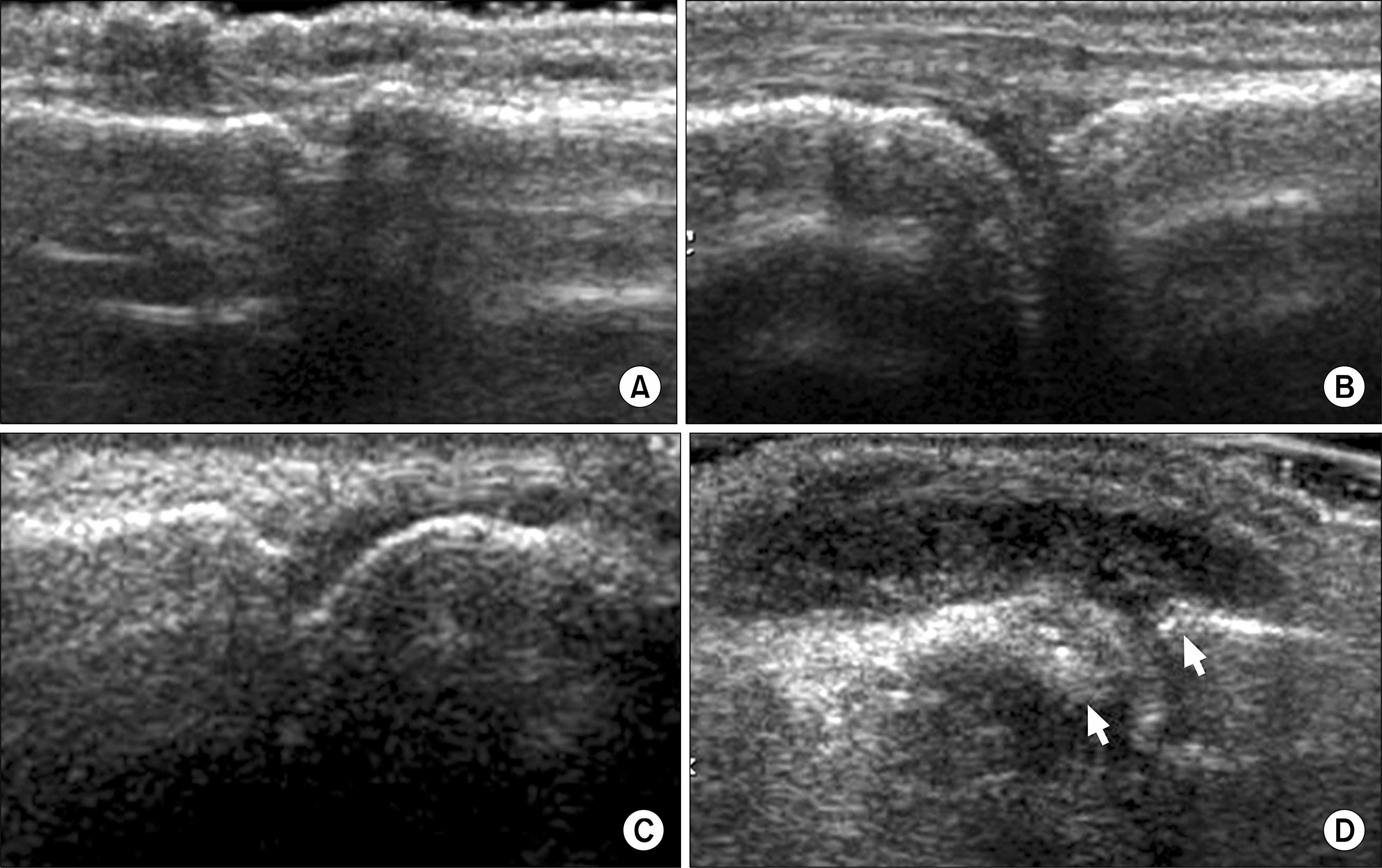
Fig. 2. Ultrasonographic findings of joint effusion can be graded according to the amount. (A) Grade 0 (no effusion), (B) grade 1 (minimal amount of fluid), (C) grade 2 (moderate amount of fluid without distension of the joint capsule), and (D) grade 3: extensive amount of fluid with distension of the joint capsule.

Fig. 3. Ultrasonographic findings of synovial proliferation. (A) Olecrenon fossa of the elbow, (B) lateral recess of the knee, (C) metacarpophalangeal joint, and (D) dorsal carpal joint.
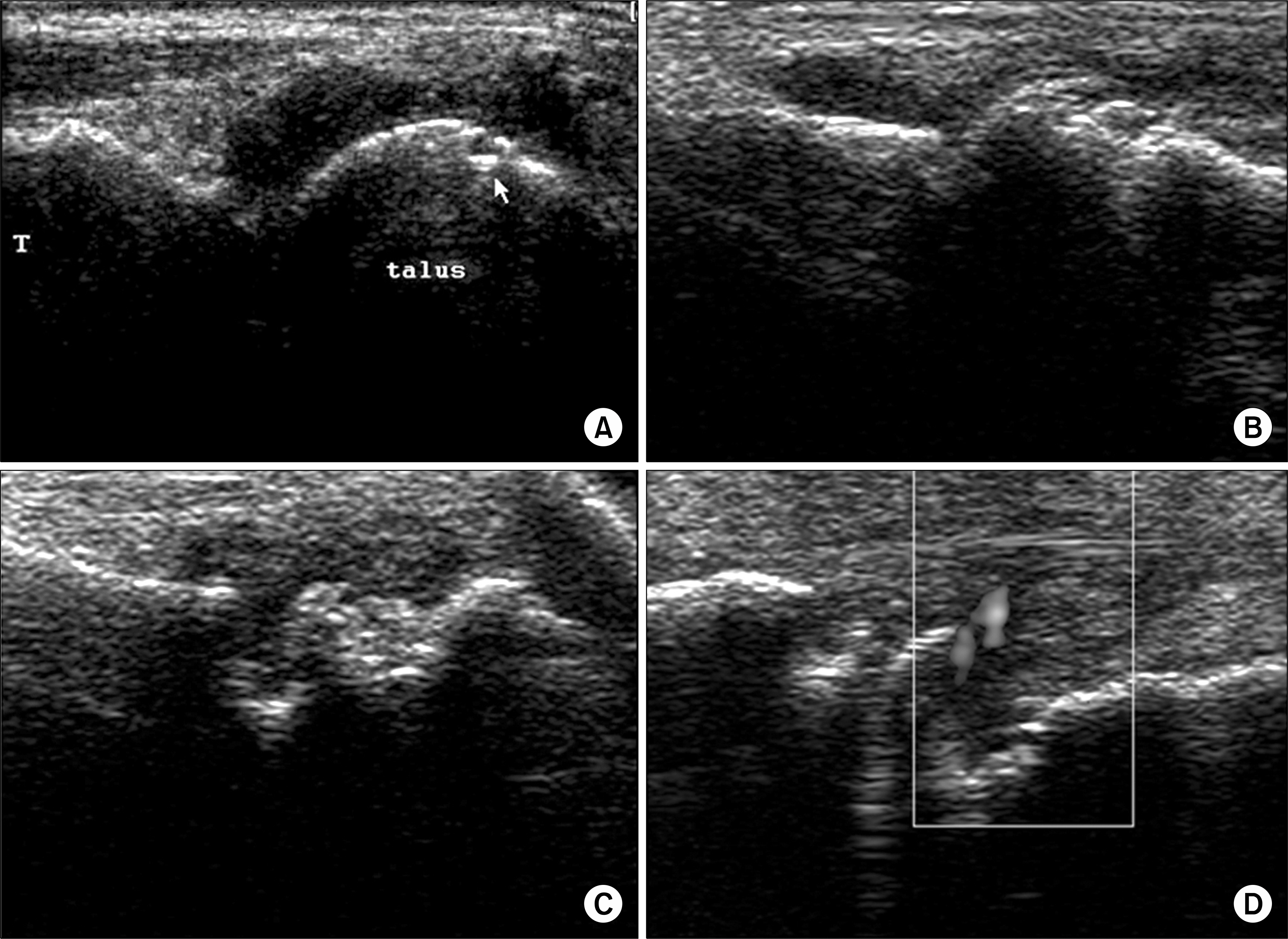
Fig. 4. Ultrasonographic findings of bone erosion. (A) Small erosion in the talus, (B) irregular and moderate-sized erosions of the metacarpal head, (C) large and deep erosion of the metacarpal head, and (D) inflammatory pannus with increased vascularity on power Doppler ultrasound in the metatarsal joint.

Fig. 5. Ultrasonographic findings of tenosynovitis. (A, B) Anechoic fluid accumulation in the sheath of posterior tibial tendon (longitudinal and transverse scans). (C, D) Hyperechoic synovial proliferation in the sheath of the extensor digitorum tendons (longitudinal and transverse scans).
Reference
-
References
1. Harrison BJ, Symmons DP, Barrett EM, Silman AJ. The performance of the 1987 ARA classification criteria for rheumatoid arthritis in a population based cohort of patients with early inflammatory polyarthritis. American Rheumatism Association. J Rheumatol. 1998; 25:2324–30.2. Chan KW, Felson DT, Yood RA, Walker AM. The lag time between onset of symptoms and diagnosis of rheumatoid arthritis. Arthritis Rheum. 1994; 37:814–20.
Article3. van Dongen H, van Aken J, Lard LR, Visser K, Ronday HK, Hulsmans HM, et al. Efficacy of methotrexate treatment in patients with probable rheumatoid arthritis: a double-blind, randomized, placebocontrolled trial. Arthritis Rheum. 2007; 56:1424–32.
Article4. Taylor PC, Steuer A, Gruber J, Cosgrove DO, Blom-ley MJ, Marsters PA, et al. Comparison of ultrasonographic assessment of synovitis and joint vascularity with radiographic evaluation in a randomized, placebocontrolled study of infliximab therapy in early rheumatoid arthritis. Arthritis Rheum. 2004; 50:1107–16.
Article5. Szkudlarek M, Narvestad E, Klarlund M, Court-Payen M, Thomsen HS, Ostergaard M. Ultrasonography of the metatarsophalangeal joints in rheumatoid arthritis: comparison with magnetic resonance imaging, conventional radiography, and clinical examination. Arthritis Rheum. 2004; 50:2103–12.
Article6. Wakefield RJ, Gibbon WW, Conaghan PG, O'Connor P, McGonagle D, Pease C, et al. The value of sonography in the detection of bone erosions in patients with rheumatoid arthritis: a comparison with conventional radiography. Arthritis Rheum. 2000; 43:2762–70.
Article7. Schmidt WA. Value of sonography in diagnosis of rheumatoid arthritis. Lancet. 2001; 357:1056–7.
Article8. Filippucci E, Iagnocco A, Meenagh G, Riente L, Delle Sedie A, Bombardieri S, et al. Ultrasound imaging for the rheumatologist VII. Ultrasound imaging in rheumatoid arthritis. Clin Exp Rheumatol. 2007; 25:5–10.9. Backhaus M, Kamradt T, Sandrock D, Loreck D, Fritz J, Wolf KJ, et al. Arthritis of the finger joints: a comprehensive approach comparing conventional radiography, scintigraphy, ultrasound, and contrast-enhanced magnetic resonance imaging. Arthritis Rheum. 1999; 42:1232–45.
Article10. Kane D, Balint PV, Sturrock RD. Ultrasonography is superior to clinical examination in the detection and localization of knee joint effusion in rheumatoid arthritis. J Rheumatol. 2003; 30:966–71.11. Wakefield RJ, Green MJ, Marzo-Ortega H, Conaghan PG, Gibbon WW, McGonagle D, et al. Should oligoarthritis be reclassified? Ultrasound reveals a high prevalence of subclinical disease. Ann Rheum Dis. 2004; 63:382–5.
Article12. Joshua F, Edmonds J, Lassere M. Power Doppler ultrasound in musculoskeletal disease: a systematic review. Semin Arthritis Rheum. 2006; 36:99–108.
Article13. Naredo E, Moller I, Cruz A, Carmona L, Garrido J. Power Doppler ultrasonographic monitoring of response to antitumor necrosis factor therapy in patients with rheumatoid arthritis. Arthritis Rheum. 2008; 58:2248–56.
Article14. Naredo E, Bonilla G, Gamero F, Uson J, Carmona L, Laffon A. Assessment of inflammatory activity in rheumatoid arthritis: a comparative study of clinical evaluation with grey scale and power Doppler ultrasonography. Ann Rheum Dis. 2005; 64:375–81.
Article15. Hau M, Kneitz C, Tony HP, Keberle M, Jahns R, Jenett M. High resolution ultrasound detects a decrease in pannus vascularisation of small finger joints in patients with rheumatoid arthritis receiving treatment with soluble tumour necrosis factor alpha receptor (etanercept). Ann Rheum Dis. 2002; 61:55–8.
Article16. Iagnocco A, Filippucci E, Perella C, Ceccarelli F, Cassara E, Alessandri C, et al. Clinical and ultrasonographic monitoring of response to adalimumab treatment in rheumatoid arthritis. J Rheumatol. 2008; 35:35–40.17. Szkudlarek M, Court-Payen M, Jacobsen S, Klarlund M, Thomsen HS, Ostergaard M. Interobserver agreement in ultrasonography of the finger and toe joints in rheumatoid arthritis. Arthritis Rheum. 2003; 48:955–62.
Article18. Pincus T. Limitations of a quantitative swollen and tender joint count to assess and monitor patients with rheumatoid arthritis. Bull NYU Hosp Jt Dis. 2008; 66:216–23.19. Wiell C, Szkudlarek M, Hasselquist M, Moller JM, Vestergaard A, Norregaard J, et al. Ultrasonography, magnetic resonance imaging, radiography, and clinical assessment of inflammatory and destructive changes in fingers and toes of patients with psoriatic arthritis. Arthritis Res Ther. 2007; 9:119.
Article20. Brown AK, Quinn MA, Karim Z, Conaghan PG, Peterfy CG, Hensor E, et al. Presence of significant synovitis in rheumatoid arthritis patients with disease-modifying antirheumatic drug-induced clinical remission: evidence from an imaging study may explain structural progression. Arthritis Rheum. 2006; 54:3761–73.
Article21. Farrant JM, O'Connor PJ, Grainger AJ. Advanced imaging in rheumatoid arthritis. Part 1: synovitis. Skeletal Radiol. 2007; 36:269–79.22. Wakefield RJ, Balint PV, Szkudlarek M, Filippucci E, Backhaus M, D'Agostino MA, et al. Musculoskeletal ultrasound including definitions for ultrasonographic pathology. J Rheumatol. 2005; 32:2485–7.23. Raza K, Lee CY, Pilling D, Heaton S, Situnayake RD, Carruthers DM, et al. Ultrasound guidance allows accurate needle placement and aspiration from small joints in patients with early inflammatory arthritis. Rheumatology (Oxford). 2003; 42:976–9.
Article24. Hoving JL, Buchbinder R, Hall S, Lawler G, Coombs P, McNealy S, et al. A comparison of magnetic resonance imaging, sonography, and radiography of the hand in patients with early rheumatoid arthritis. J Rheumatol. 2004; 31:663–75.25. Magnani M, Salizzoni E, Mule R, Fusconi M, Meliconi R, Galletti S. Ultrasonography detection of early bone erosions in the metacarpophalangeal joints of patients with rheumatoid arthritis. Clin Exp Rheumatol. 2004; 22:743–8.26. Taylor PC. The value of sensitive imaging modalities in rheumatoid arthritis. Arthritis Res Ther. 2003; 5:210–3.27. Newman JS, Adler RS, Bude RO, Rubin JM. Detection of soft-tissue hyperemia: value of power Doppler sonography. AJR Am J Roentgenol. 1994; 163:385–9.
Article28. Walther M, Harms H, Krenn V, Radke S, Faehndrich TP, Gohlke F. Correlation of power Doppler sonography with vascularity of the synovial tissue of the knee joint in patients with osteoarthritis and rheumatoid arthritis. Arthritis Rheum. 2001; 44:331–8.
Article29. Taylor PC. Serum vascular markers and vascular imaging in assessment of rheumatoid arthritis disease activity and response to therapy. Rheumatology (Oxford). 2005; 44:721–8.
Article30. Fiocco U, Cozzi L, Rubaltelli L, Rigon C, De Candia A, Tregnaghi A, et al. Long-term sonographic followup of rheumatoid and psoriatic proliferative knee joint synovitis. Br J Rheumatol. 1996; 35:155–63.
Article31. Schmidt WA, Volker L, Zacher J, Schlafke M, Ruhnke M, Gromnica-Ihle E. Colour Doppler ultrasonography to detect pannus in knee joint synovitis. Clin Exp Rheumatol. 2000; 18:439–44.32. Qvistgaard E, Rogind H, Torp-Pedersen S, Terslev L, Danneskiold-Samsoe B, Bliddal H. Quantitative ultrasonography in rheumatoid arthritis: evaluation of inflammation by Doppler technique. Ann Rheum Dis. 2001; 60:690–3.33. Naredo E, Rodriguez M, Campos C, Rodriguez-Heredia JM, Medina JA, Giner E, et al. Validity, reproducibility, and responsiveness of a twelve-joint simplified power doppler ultrasonographic assessment of joint inflammation in rheumatoid arthritis. Arthritis Rheum. 2008; 59:515–22.
Article34. Newman JS, Laing TJ, McCarthy CJ, Adler RS. Power Doppler sonography of synovitis: assessment of therapeutic response-preliminary observations. Radiology. 1996; 198:582–4.
Article35. Filippucci E, Farina A, Carotti M, Salaffi F, Grassi W. Grey scale and power Doppler sonographic changes induced by intraarticular steroid injection treatment. Ann Rheum Dis. 2004; 63:740–3.
Article36. Salaffi F, Carotti M, Manganelli P, Filippucci E, Giuseppetti GM, Grassi W. Contrast-enhanced power Doppler sonography of knee synovitis in rheumatoid arthritis: assessment of therapeutic response. Clin Rheumatol. 2004; 23:285–90.
Article37. Ribbens C, Andre B, Marcelis S, Kaye O, Mathy L, Bonnet V, et al. Rheumatoid hand joint synovitis: gray-scale and power Doppler US quantifications following antitumor necrosis factor-alpha treatment: pilot study. Radiology. 2003; 229:562–9.38. Fiocco U, Ferro F, Vezzu M, Cozzi L, Checchetto C, Sfriso P, et al. Rheumatoid and psoriatic knee synovitis: clinical, grey scale, and power Doppler ultrasound assessment of the response to etanercept. Ann Rheum Dis. 2005; 64:899–905.
Article39. Grassi W, Filippucci E. Is power Doppler sonography the new frontier in therapy monitoring? Clin Exp Rheumatol. 2003; 21:424–8.40. Kane D, Balint PV, Sturrock R, Grassi W. Musculoskeletal ultrasound-a state of the art review in rheumatology. Part 1: current controversies and issues in the development of musculoskeletal ultrasound in rheumatology. Rheumatology (Oxford). 2004; 43:823–8.
Article41. Kubassova O, Boesen M, Peloschek P, Langs G, Cimmino MA, Bliddal H, et al. Quantifying disease activity and damage by imaging in rheumatoid arthritis and osteoarthritis. Ann N Y Acad Sci. 2009; 1154:207–38.
Article42. Boutry N, Carmo CC, Flipo RM, Cotten A. Early rheumatoid arthritis and its differentiation from other joint abnormalities. Eur J Radiol. 2009. [Epub ahead of print].
Article43. Pierre-Jerome C, Bekkelund SI, Mellgren SI, Torberg-sen T, Husby G, Nordstrom R. The rheumatoid wrist: bilateral MR analysis of the distribution of rheumatoid lesions in axial plan in a female population. Clin Rheumatol. 1997; 16:80–6.
Article44. Alasaarela E, Suramo I, Tervonen O, Lahde S, Takalo R, Hakala M. Evaluation of humeral head erosions in rheumatoid arthritis: a comparison of ultrasonography, magnetic resonance imaging, computed tomography and plain radiography. Br J Rheumatol. 1998; 37.
Article45. Iagnocco A, Coari G, Zoppini A. Sonographic evaluation of femoral condylar cartilage in osteoarthritis and rheumatoid arthritis. Scand J Rheumatol. 1992; 21:201–3.
Article46. Moller B, Bonel H, Rotzetter M, Villiger PM, Ziswiler HR. Measuring finger joint cartilage by ultrasound as a promising alternative to conventional radiograph imaging. Arthritis Rheum. 2009; 61:435–41.
Article47. Eshed I, Feist E, Althoff CE, Hamm B, Konen E, Burmester GR, et al. Tenosynovitis of the flexor tendons of the hand detected by MRI: an early indicator of rheumatoid arthritis. Rheumatology (Oxford). 2009. [Epub ahead of print].
Article48. Boutry N, Morel M, Flipo RM, Demondion X, Cotten A. Early rheumatoid arthritis: a review of MRI and sonographic findings. AJR Am J Roentgenol. 2007; 189:1502–9.
Article49. Hameed B, Pilcher J, Heron C, Kiely PD. The relation between composite ultrasound measures and the DAS28 score, its components and acute phase markers in adult RA. Rheumatology (Oxford). 2008; 47:476–80.
Article50. Ostergaard M, Ejbjerg B, Szkudlarek M. Imaging in early rheumatoid arthritis: roles of magnetic resonance imaging, ultrasonography, conventional radiography and computed tomography. Best Pract Res Clin Rheumatol. 2005; 19:91–116.
Article51. Downey DB, Fenster A, Williams JC. Clinical utility of three-dimensional US. Radiographics. 2000; 20:559–71.
Article52. Filippucci E, Meenagh G, Epis O, Iagnocco A, Riente L, Delle Sedie A, et al. Ultrasound imaging for the rheumatologist. XIII. New trends. Three-dimensional ultrasonography. Clin Exp Rheumatol. 2008; 26:1–4.53. Strunk J, Strube K, Rumbaur C, Lange U, Muller-Ladner U. Interobserver agreement in two- and three-dimensional power Doppler sonographic assessment of synovial vascularity during antiinflammatory treatment in patients with rheumatoid arthritis. Ultraschall Med. 2007; 28:409–15.
Article54. Strunk J, Strube K, Muller-Ladner U, Lange U. Three dimensional power Doppler ultrasonography confirms early reduction of synovial perfusion after intraarticular steroid injection. Ann Rheum Dis. 2006; 65:411–2.
Article55. Naredo E, Moller I, Moragues C, de Agustin JJ, Scheel AK, Grassi W, et al. Interobserver reliability in musculoskeletal ultrasonography: results from a “Teach the Teachers” rheumatologist course. Ann Rheum Dis. 2006; 65:14–9.
Article56. Scheel AK, Schmidt WA, Hermann KG, Bruyn GA, D'Agostino MA, Grassi W, et al. Interobserver reliability of rheumatologists performing musculoskeletal ultrasonography: results from a EULAR “Train the trainers” course. Ann Rheum Dis. 2005; 64:1043–9.
Article57. Terslev L, Torp-Pedersen S, Savnik A, von der Recke P, Qvistgaard E, Danneskiold-Samsoe B, et al. Doppler ultrasound and magnetic resonance imaging of synovial inflammation of the hand in rheumatoid arthritis: a comparative study. Arthritis Rheum. 2003; 48:2434–41.
Article58. Scheel AK, Hermann KG, Kahler E, Pasewaldt D, Fritz J, Hamm B, et al. A novel ultrasonographic synovitis scoring system suitable for analyzing finger joint inflammation in rheumatoid arthritis. Arthritis Rheum. 2005; 52:733–43.
Article59. Naredo E, Gamero F, Bonilla G, Uson J, Carmona L, Laffon A. Ultrasonographic assessment of inflammatory activity in rheumatoid arthritis: comparison of extended versus reduced joint evaluation. Clin Exp Rheumatol. 2005; 23:881–4.60. Weidekamm C, Koller M, Weber M, Kainberger F. Diagnostic value of high-resolution B-mode and doppler sonography for imaging of hand and finger joints in rheumatoid arthritis. Arthritis Rheum. 2003; 48:325–33.
Article61. Strunk J, Heinemann E, Neeck G, Schmidt KL, Lange U. A new approach to studying angiogenesis in rheumatoid arthritis by means of power Doppler ultrasonography and measurement of serum vascular endothelial growth factor. Rheumatology (Oxford). 2004; 43:1480–3.
Article
