Extensive gingival necrosis and sequestration of the alveolar bone caused by methimazole-induced neutropenia and three-year follow-up
- Affiliations
-
- 1Department of Oral and Maxillofacial Pathology, School of Dentistry and Research Center for Tooth & Periodontal Regeneration (MRC), Kyung Hee University, Seoul, Korea.
- 2Department of Periodontology, School of Dentistry, Kyung Hee University, Seoul, Korea. periokkl@khu.ac.kr
- 3Department of Periodontics, Kyung Hee University Dental Hospital at Gangdong, Seoul, Korea.
- 4Institute of Oral Biology, School of Dentistry, Kyung Hee University, Seoul, Korea.
- KMID: 2329659
- DOI: http://doi.org/10.5051/jpis.2015.45.2.76
Abstract
- PURPOSE
Methimazole is an anti-thyroid drug that can cause life-threatening neutropenia in rare situations. The aim of this case report is to describe a set of oral complications associated with methimazole-induced neutropenia and the healing of the gingiva after proper treatment.
METHODS
A 31-year-old female patient hospitalized for systemic symptoms of sore throat and fever and showing extensive gingival necrosis with pain was referred to the Department of Periodontics from the Department of Endocrinology. Methimazole-induced neutropenia was diagnosed based on blood test results and her medical history. Methimazole was discontinued and a range of treatments was administered, including the injection of granulocyte colony stimulating factor.
RESULTS
After systemic treatment, the gingiva began to heal as the neutrophil count increased. Approximately one year later, the gingiva had returned to a normal appearance. Twenty-one months after treatment, sequestra of the alveolar bone that had broken through the gingiva were removed. Periodic supportive periodontal treatment has been continued uneventfully.
CONCLUSIONS
The oral manifestations of gingival necrosis and ulcerations, in combination with systemic symptoms such as fever and sore throat, are the critical signs presented in the early stages of drug-induced neutropenia. Therefore, dentists need to be aware of these oral complications in order to make an accurate diagnosis and to ensure that prompt medical intervention is provided.
Keyword
MeSH Terms
Figure
-
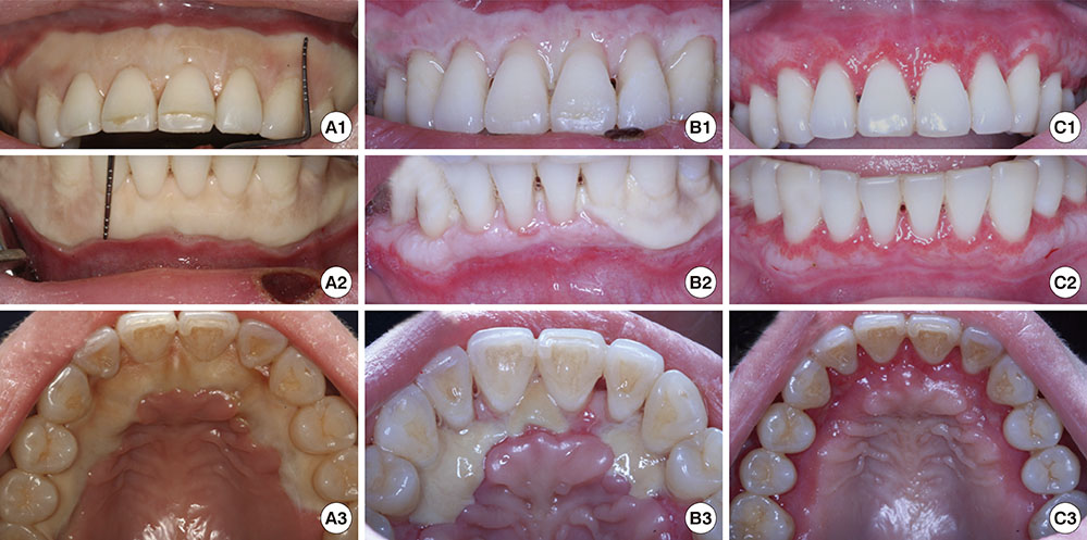
Figure 1 At the first dental visit (A1, A2, A3), a wide zone of whitish necrotic lesions was found in the gingiva, extending across almost the entire dentition. An erythematous margin limiting the necrotic gingival zone was formed at the alveolar mucosa. A crust was present on the left side of the lower lip. Five days after the first dental visit (B1, B2, B3), the necrotic area decreased remarkably and re-epithelialization had started. Two weeks after the first dental visit (C1, C2, C3), pale and pink gingiva, reddish gingival margins, and exposed root surfaces were observed.

Figure 2 Eight weeks after the first dental visit (A), the gingiva was reddish-pink and much more epithelialized. At 18 weeks of follow-up (B), the gingival margins were re-epithelialized and the size of the interdental spaces had decreased. At a nine-month follow-up (C), the embrasure spaces were almost completely filled with gingival tissue, even though some calculus and plaque were present. At one year of follow-up (D), the upper and lower gingiva showed a normal appearance.

Figure 3 Necrotic alveolar bones protruding out of the marginal gingiva (arrow) were found around the maxillary left central incisor and canine at a 21-month follow-up visit (A, C) and the sequestra of the bone were removed (B, D, E). At a three-year follow-up visit, the gingiva were found to have a normal appearance (F).
Reference
-
1. Tamai H, Mukuta T, Matsubayashi S, Fukata S, Komaki G, Kuma K, et al. Treatment of methimazole-induced agranulocytosis using recombinant human granulocyte colony-stimulating factor (rhG-CSF). J Clin Endocrinol Metab. 1993; 77:1356–1360.
Article2. Dai WX, Zhang JD, Zhan SW, Xu BZ, Jin H, Yao Y, et al. Retrospective analysis of 18 cases of antithyroid drug (ATD)-induced agranulocytosis. Endocr J. 2002; 49:29–33.
Article3. Chen DF, Chao IM, Huang SH. Neutropenic colitis with cecal perforation during antithyroid therapy. J Formos Med Assoc. 2003; 102:644–646.4. Jabr FI. Methimazole-induced severe febrile neutropenia responding to recombinant human granulocyte colony stimulating factor. South Med J. 2008; 101:665.
Article5. Meyer-Gessner M, Benker G, Lederbogen S, Olbricht T, Reinwein D. Antithyroid drug-induced agranulocytosis: clinical experience with ten patients treated at one institution and review of the literature. J Endocrinol Invest. 1994; 17:29–36.
Article6. Hou GL, Tsai CC. Oral manifestations of agranulocytosis associated with methimazole therapy. J Periodontol. 1988; 59:244–248.
Article7. Andersohn F, Konzen C, Garbe E. Systematic review: agranulocytosis induced by nonchemotherapy drugs. Ann Intern Med. 2007; 146:657–665.
Article8. Miller DR, Lamster IB, Chasens AI. Role of the polymorphonuclear leukocyte in periodontal health and disease. J Clin Periodontol. 1984; 11:1–15.
Article9. Goultschin J, Attal U, Goldstein M, Boyan BD, Schwartz Z. The relationship between peripheral levels of leukocytes and neutrophils and periodontal disease status in a patient with congenital neutropenia. J Periodontol. 2000; 71:1499–1505.
Article10. Tajiri J, Noguchi S, Murakami T, Murakami N. Antithyroid drug-induced agranulocytosis. The usefulness of routine white blood cell count monitoring. Arch Intern Med. 1990; 150:621–624.
Article11. Chien MN, Wang CH, Tsan KW. Methimazole-induced agranulocytosis treated with recombinant human granulocyte colony-stimulating factor (rhG-CSF): a case report. Zhonghua Yi Xue Za Zhi (Taipei). 1995; 56:351–355.12. Espinosa DJ. Analytical review of multicenter studies with polycresulene for hemorrhoidal pathologies. Acta Gastroenterol Latinoam. 2000; 30:177–186.13. Gunsolley JC. A meta-analysis of six-month studies of antiplaque and antigingivitis agents. J Am Dent Assoc. 2006; 137:1649–1657.
Article
- Full Text Links
-
- Actions
-
Cited
- CITED
-
- Close
- Share
-
- Similar articles
-
- Mandibular bone necrosis after use of paraformaldehyde-containing paste
- A Case of Acute Appendicitis in a Patient with Methimazole-Induced Agranulocytosis
- A Case of Methimazole-Induced Cholestatic Jaundice with Steroid Therapy
- Cytoplasmic Anti-Neutrophil Cytoplasmic Antibody Positive Diffuse Alveolar Hemorrhage Associated with Methimazole
- A case of Methimazole-Induced Cholestatic Jaundice With Agranulocytosis
