Ultrasonographic Measurement of the Thickness of Axillary Recess Capsule in Healthy Volunteers
- Affiliations
-
- 1Department of Rehabilitation Medicine, Keimyung University School of Medicine, Daegu, Korea. ri-pheonix@hanmail.net
- 2Pain Research Center, Keimyung University School of Medicine, Daegu, Korea.
- 3Institute for Medical Science, Keimyung University School of Medicine, Daegu, Korea.
- KMID: 2327613
- DOI: http://doi.org/10.5535/arm.2016.40.3.502
Abstract
OBJECTIVE
To evaluate the inter-rater and intra-rater reliability of ultrasonographic measurements of axillary recess (AR) thickness in healthy individuals, and to analyze the factors affecting the thickness of the AR capsule.
METHODS
We recruited 20 healthy individuals (10 male, 10 female) with a mean age of 37 years (standard deviation ±10). Two physiatrists (an experienced and a novice rater) independently investigated the AR thickness in three rounds. The AR thickness was measured for each individual at three shoulder abduction angles (50°, 70°, and 90°). Intra-class correlation (ICC) coefficients were used to assess the reproducibility of each measurement.
RESULTS
Excellent intra-rater reliability coefficients were observed at the three shoulder abduction angles, in the analysis of both raters. The inter-rater reliability coefficient was also was excellent in both studies. There were significant differences in the AR thickness, according to the angle of shoulder abduction. The AR was thicker at 50° than at 70° and 90° (all p<0.001), and the AR was thicker at 70° than at 90° (p<0.001). Height (r=0.62, p=0.003) and body mass index (r=0.52, p=0.019) were positively correlated with AR thickness. Males had a thicker AR capsule than females at all three angles (all p<0.001).
CONCLUSION
Ultrasonographic measurements of AR thickness in healthy individuals demonstrate excellent intra-rater and inter-rater reliability. AR thickness may depend on anthropometric variables and position of the shoulder.
MeSH Terms
Figure
-
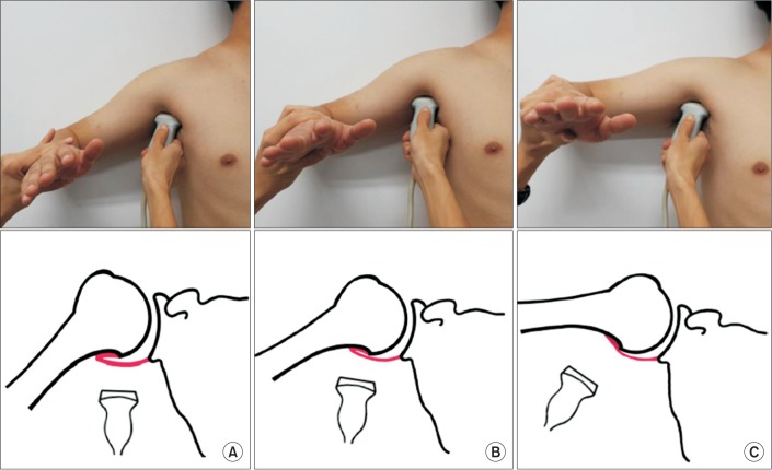
Fig. 1 Evaluation of the axillary recess capsule, using ultrasonography and schematic drawings, at three different positions according to the degrees of shoulder abduction (50°, 70°, and 90°).
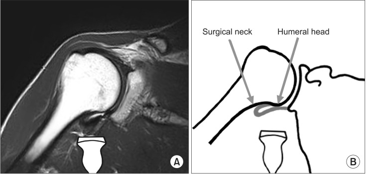
Fig. 2 (A) Coronal view of shoulder magnetic resonance imaging showing the region of interest in the ultrasonographic measurement of axillary recess thickness. (B) Schematic illustration of axillary recess.
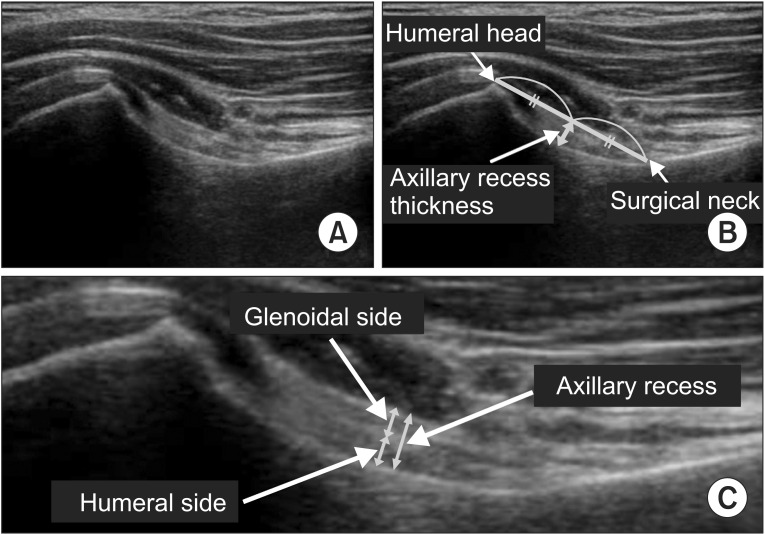
Fig. 3 (A) Axillary recess (AR) capsule on a frozen image. (B) Determination of the thickness of the AR capsule. (C) Humeral and glenoid sides of the AR capsule. The AR thickness is defined as the total summation of each thickness of the glenoid and humeral capsules.
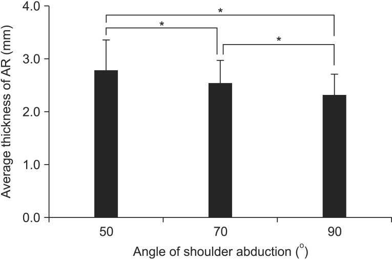
Fig. 4 Axillary recess (AR) thickness according to three different positions. *p<0.001.
Reference
-
1. Hannafin JA, Chiaia TA. Adhesive capsulitis: a treatment approach. Clin Orthop Relat Res. 2000; 372:95–109. PMID: 10738419.2. Hsu JE, Anakwenze OA, Warrender WJ, Abboud JA. Current review of adhesive capsulitis. J Shoulder Elbow Surg. 2011; 20:502–514. PMID: 21167743.
Article3. Lee MH, Ahn JM, Muhle C, Kim SH, Park JS, Kim SH, et al. Adhesive capsulitis of the shoulder: diagnosis using magnetic resonance arthrography, with arthroscopic findings as the standard. J Comput Assist Tomogr. 2003; 27:901–906. PMID: 14600458.4. Manton GL, Schweitzer ME, Weishaupt D, Karasick D. Utility of MR arthrography in the diagnosis of adhesive capsulitis. Skeletal Radiol. 2001; 30:326–330. PMID: 11465773.
Article5. Mengiardi B, Pfirrmann CW, Gerber C, Hodler J, Zanetti M. Frozen shoulder: MR arthrographic findings. Radiology. 2004; 233:486–492. PMID: 15358849.
Article6. Sofka CM, Ciavarra GA, Hannafin JA, Cordasco FA, Potter HG. Magnetic resonance imaging of adhesive capsulitis: correlation with clinical staging. HSS J. 2008; 4:164–169. PMID: 18815860.
Article7. Song KD, Kwon JW, Yoon YC, Choi SH. Indirect MR arthrographic findings of adhesive capsulitis. AJR Am J Roentgenol. 2011; 197:W1105–W1109. PMID: 22109326.
Article8. Tamai K, Akutsu M, Yano Y. Primary frozen shoulder: brief review of pathology and imaging abnormalities. J Orthop Sci. 2014; 19:1–5. PMID: 24306579.
Article9. Tamai K, Yamato M. Abnormal synovium in the frozen shoulder: a preliminary report with dynamic magnetic resonance imaging. J Shoulder Elbow Surg. 1997; 6:534–543. PMID: 9437603.
Article10. Lefevre-Colau MM, Drape JL, Fayad F, Rannou F, Diche T, Minvielle F, et al. Magnetic resonance imaging of shoulders with idiopathic adhesive capsulitis: reliability of measures. Eur Radiol. 2005; 15:2415–2422. PMID: 16003508.
Article11. Ryu KN, Lee SW, Rhee YG, Lim JH. Adhesive capsulitis of the shoulder joint: usefulness of dynamic sonography. J Ultrasound Med. 1993; 12:445–449. PMID: 8411327.
Article12. Walmsley S, Osmotherly PG, Walker CJ, Rivett DA. Power Doppler ultrasonography in the early diagnosis of primary/idiopathic adhesive capsulitis: an exploratory study. J Manipulative Physiol Ther. 2013; 36:428–435. PMID: 23830711.
Article13. Dasgupta B, Cimmino MA, Kremers HM, Schmidt WA, Schirmer M, Salvarani C, et al. 2012 Provisional classification criteria for polymyalgia rheumatica: a European League Against Rheumatism/American College of Rheumatology collaborative initiative. Arthritis Rheum. 2012; 64:943–954. PMID: 22389040.
Article14. Ruta S, Rosa J, Navarta DA, Saucedo C, Catoggio LJ, Monaco RG, et al. Ultrasound assessment of new onset bilateral painful shoulder in patients with polymyalgia rheumatica and rheumatoid arthritis. Clin Rheumatol. 2012; 31:1383–1387. PMID: 22684205.
Article15. Walter SD, Eliasziw M, Donner A. Sample size and optimal designs for reliability studies. Stat Med. 1998; 17:101–110. PMID: 9463853.
Article16. Shrout PE, Fleiss JL. Intraclass correlations: uses in assessing rater reliability. Psychol Bull. 1979; 86:420–428. PMID: 18839484.
Article17. Codman E. The shoulder: rupture of the supraspinatus tendon and other lesions in or about the subacromial bursa. Boston: T. Todd Company;1934.18. Sher JS, Uribe JW, Posada A, Murphy BJ, Zlatkin MB. Abnormal findings on magnetic resonance images of asymptomatic shoulders. J Bone Joint Surg Am. 1995; 77:10–15. PMID: 7822341.
Article19. Jung JY, Jee WH, Chun HJ, Kim YS, Chung YG, Kim JM. Adhesive capsulitis of the shoulder: evaluation with MR arthrography. Eur Radiol. 2006; 16:791–796. PMID: 16228212.
Article20. Kim du H, Sung DH, Ga HY, Choi JY. Metabolic patterns of the shoulder joint on (18)F-fluorodeoxyglucose positron emission tomography/computed tomography in adhesive capsulitis. Ann Nucl Med. 2014; 28:136–144. PMID: 24317879.
Article21. Kim I, Yi JH, Lee J, Bae JH, Lim JK, Yoon JP, et al. Limited subacromial gliding of the supraspinatus tendon during dynamic ultrasonography can predict a decrease in capacity and MR arthrographic features of the shoulder joint. Eur Radiol. 2012; 22:2365–2370. PMID: 22898934.
Article22. Lee JC, Sykes C, Saifuddin A, Connell D. Adhesive capsulitis: sonographic changes in the rotator cuff interval with arthroscopic correlation. Skeletal Radiol. 2005; 34:522–527. PMID: 15999280.
Article23. Carrillon Y, Noel E, Fantino O, Perrin-Fayolle O, Tran-Minh VA. Magnetic resonance imaging findings in idiopathic adhesive capsulitis of the shoulder. Rev Rhum Engl Ed. 1999; 66:201–206. PMID: 10339775.24. Connell D, Padmanabhan R, Buchbinder R. Adhesive capsulitis: role of MR imaging in differential diagnosis. Eur Radiol. 2002; 12:2100–2106. PMID: 12136330.
Article25. Emig EW, Schweitzer ME, Karasick D, Lubowitz J. Adhesive capsulitis of the shoulder: MR diagnosis. AJR Am J Roentgenol. 1995; 164:1457–1459. PMID: 7754892.
Article26. Rainis EJ, Maas SA, Henninger HB, McMahon PJ, Weiss JA, Debski RE. Material properties of the axillary pouch of the glenohumeral capsule: is isotropic material symmetry appropriate? J Biomech Eng. 2009; 131:031007. PMID: 19154066.
Article27. Urayama M, Itoi E, Sashi R, Minagawa H, Sato K. Capsular elongation in shoulders with recurrent anterior dislocation: quantitative assessment with magnetic resonance arthrography. Am J Sports Med. 2003; 31:64–67. PMID: 12531759.
- Full Text Links
-
- Actions
-
Cited
- CITED
-
- Close
- Share
-
- Similar articles
-
- Ultrasonographic measurement of the axillary recess thickness in an asymptomatic shoulder
- Conventioinal MRI Finding in a Case of Adhesive Shoulder Capsulitis
- Ultrasonographic and macroscopic comparison of the thickness of the capsule, corium, and soft tissues in bovine claws: an in vitro study
- Comparison of Skin Thickness in Lymphedema Using Ultrasonography and Skin Biopsy
- Measurements of heel pad thickness in normal Koreans by ultrasound and conventional x-ray
