J Korean Soc Spine Surg.
2015 Jun;22(2):55-59. 10.4184/jkss.2015.22.2.55.
Thoracic Vertebral Fracture due to Spinal Tuberculosis which was Misdiagnosed as Matastatic Cancer: A Case Report
- Affiliations
-
- 1Department of Orthopedic Surgery, Asan Medical Center, University of Ulsan College of Medicine, Seoul, Korea. doctork78@hanmail.net
- KMID: 2322953
- DOI: http://doi.org/10.4184/jkss.2015.22.2.55
Abstract
- STUDY DESIGN: A case report.
OBJECTIVES
To report the case of a patient whose preoperative imaging results seemed to show metastatic spine tumor but who actually had a vertebral pathologic fracture caused by spine tuberculosis. SUMMARY OF LITERATURE REVIEW: Tuberculosis spondylitis is classified into peridiscal, central, anterior, and posterior spondylitis according to the portion involved, and central spondylitis can be mistaken as a tumor.
MATERIALS AND METHODS
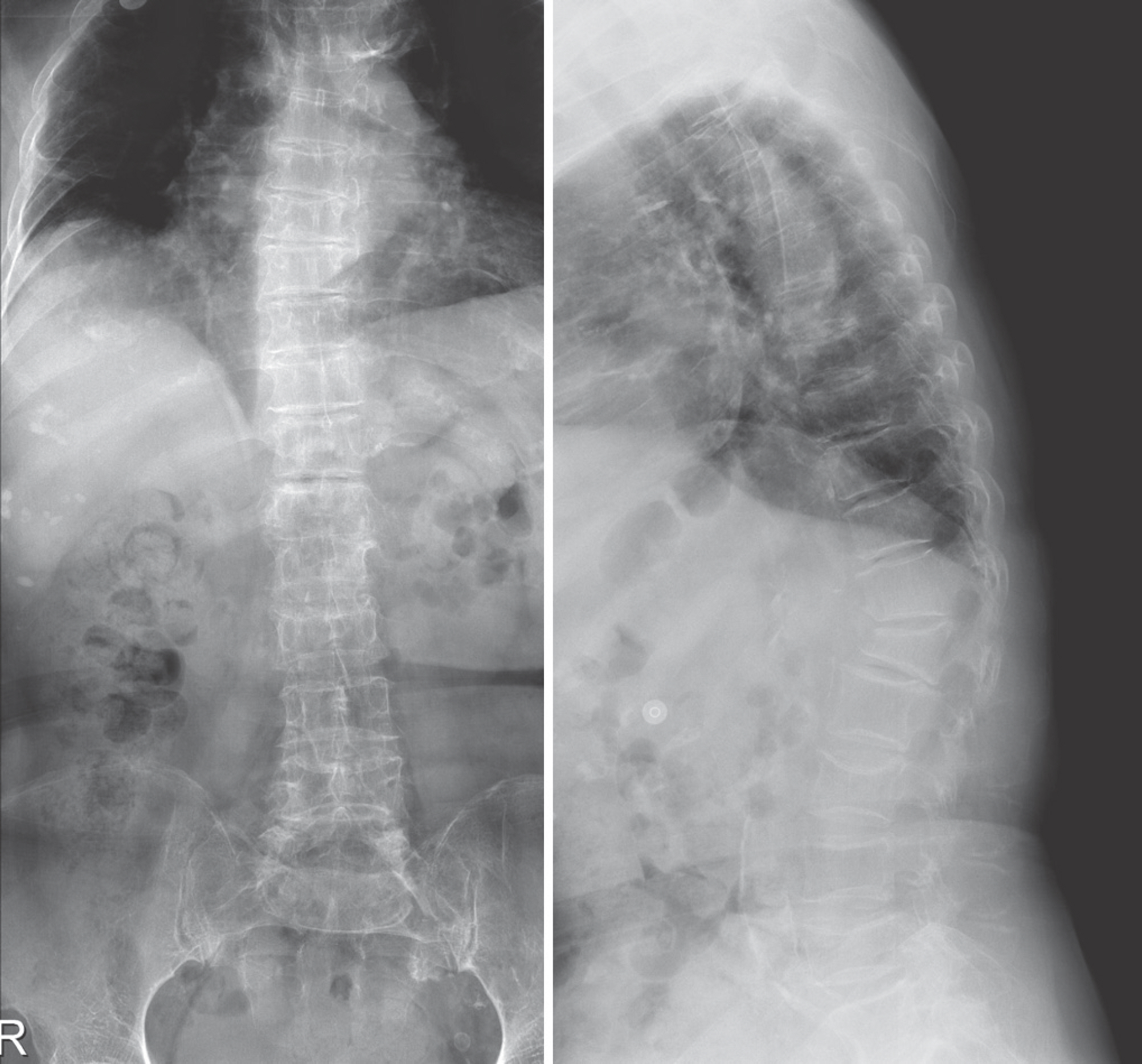
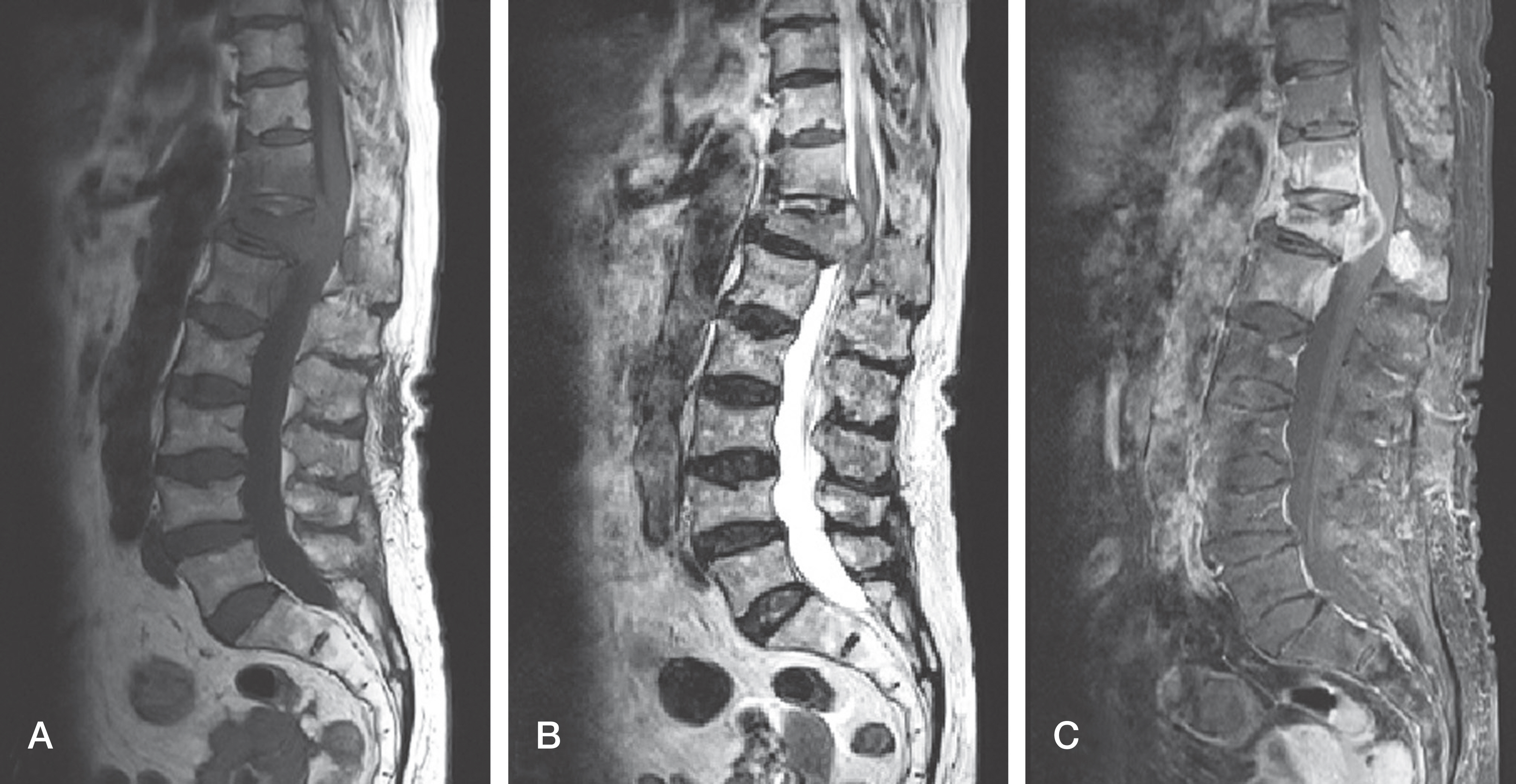
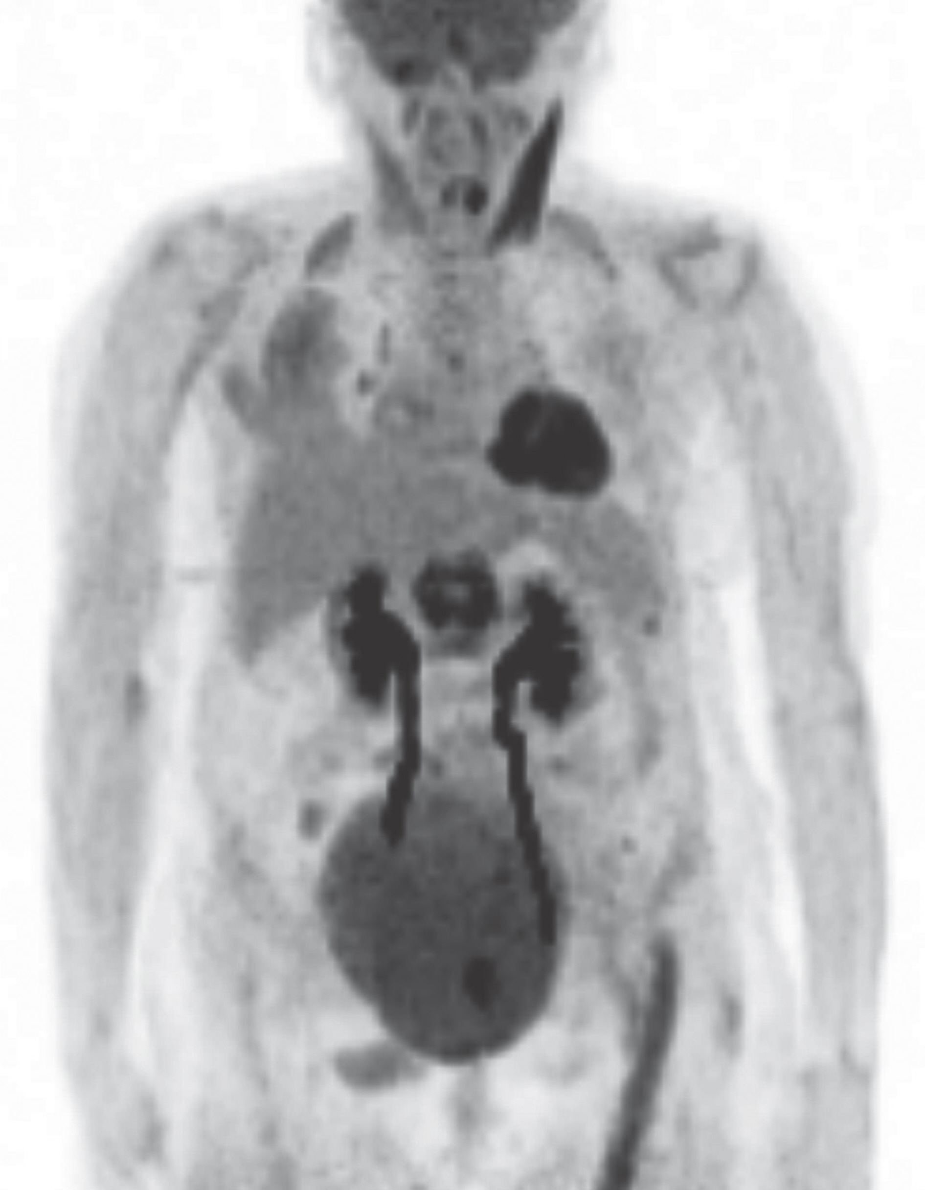
Imaging studies were performed in a 79-year-old female with progressive lower extremity weakness. We found a T12 pathologic vertebral fracture, which was suspected to be metastatic cancer.
RESULTS
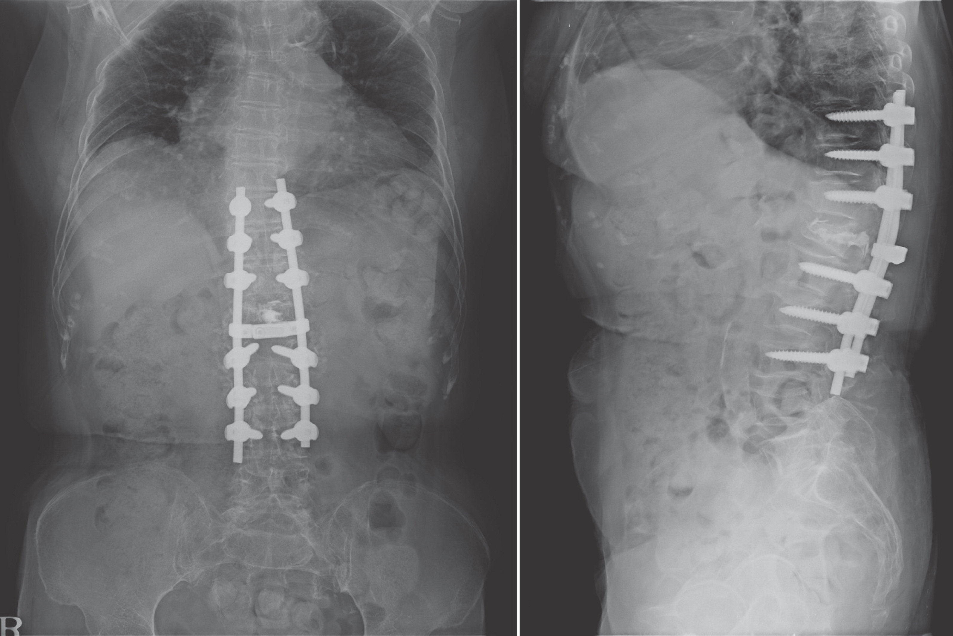
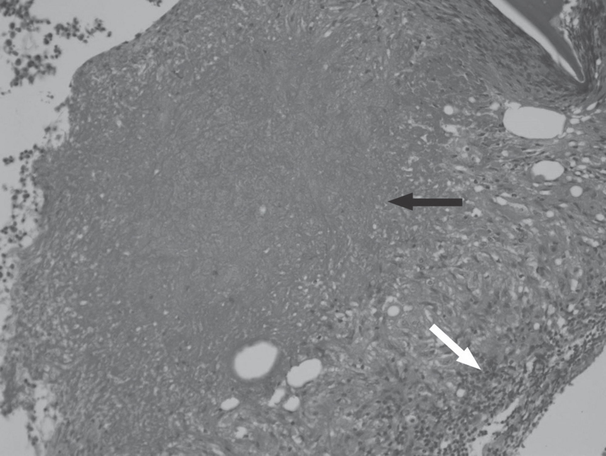
We performed surgery and found spine tuberculosis in the pathological and immunological examinations. Two weeks postoperatively, the patient could walk with crutches and underwent anti-tuberculosis therapy.
CONCLUSIONS
Even when the results of imaging studies predict spinal metastasis, we should keep in mind the possibility of spinal tuberculosis.
MeSH Terms
Figure
Reference
-
1. Joint committee for the development of guidelines for tuberculosis, prevention Kcfdca: Korea guidelines for tuberculosis first edition. 2011.2. Sankaran-Kutty M. Atypical tuberculous spondylitis. Int Orthop. 1992; 16:69–74.
Article3. Currie S, Galea-Soler S, Barron D, et al. MRI characteristics of tuberculous spondylitis. Clin Radiol. 2011; 66:778–87.
Article4. Ha KY, Na KT, Kee SR, et al. Tuberculosis of the Spine: A new Understanding of an Old Disease. J Korean Soc Spine Surg. 2014; 21:41–7.
Article5. Jahng J, Kim YH, Lee KS. Tuberculosis of the lower lumbar spine with an atypical radiological presentation - a case mimicking a malignancy. Asian Spine J. 2007; 1:102–5.6. Jeon CH, Yoon JK, Cho JH, et al. Usefulness of Fluorine-18 FDG-PET in the Diagnosis of Vertebral Pathologic Fracture. J Korean Soc Spine Surg. 2006; 13:191–9.
Article7. Golden MP, Vikram HR. Extrapulmonary tuberculosis: an overview. Am Fam Physician. 2005; 72:1761–8.8. Dosanjh DP, Hinks TS, Innes JA, et al. Improved diagnostic evaluation of suspected tuberculosis. Ann Intern Med. 2008; 148:325–36.
Article9. Pai M, Riley LW, Colford JM, et al. Interferon-gamma as-says in the immunodiagnosis of tuberculosis: a systematic review. Lancet Infect Dis. 2004; 4:761–76.10. Dewan PK, Grinsdale J, Kawamura LM. Low sensitivity of a whole-blood interferon-gamma release assay for detection of active tuberculosis. Clin Infect Dis. 2007; 44:69–73.
- Full Text Links
- Actions
-
Cited
- CITED
-
- Close
- Share
- Similar articles
-
- Guillain-Barre Syndrome Following Spinal Fusion for Thoracic Vertebral Fracture
- Tuberculosis Affecting Multiple Vertebral Bodies
- Atypical Presentation of Spinal Tuberculosis Misadiagnosed as Metastatic Spine Tumor
- Post tetanic thoracic vertebral change
- Chronic Back Pain Proven to Be Spinal Tuberculosis: A report of 2 cases

