Axillary Metastasis in the Abdominal Dermatofibrosarcoma Protuberans with the Fibrosarcomatous Changes: A Case Report
- Affiliations
-
- 1Department of Radiology, CHA Bundang Medical Center, CHA University, Seongnam, Korea. hhkkjung@hanmail.net
- 2Department of Pathology, CHA Bundang Medical Center, CHA University, Seongnam, Korea.
Abstract
- Most distant metastases of dermatofibrosarcoma protuberans (DFSP) occurs with the repeated local recurrences and mainly in the lungs. Focusing on the imaging features, we report a unique occurrence of the distant metastasis of DFSP with fibrosarcomatous changes to the axillary soft tissue without previous local recurrences, possibly misdiagnosed as the accessory breast cancer.
MeSH Terms
Figure
-

Fig. 1 A postoperative routine chest CT axial image revealed a 2 cm sized round and solid nodule with mild homogenous enhancement in the left axilla (arrow). Pulmonary metastasis was absent and no locally recurred evidences were found on chest and abdominal CT scans.

Fig. 2 Physical examination showed postoperative scar in the left abdominal skin and focal bulding with a blue skin color change in the left axilla (arrow).
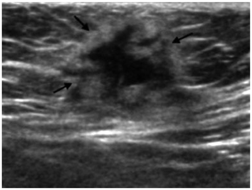
Fig. 3 A 2.1 × 1.9 cm sized irregular and hypoechoic mass with a hyperechoic rim (arrows) was seen at the left axillary subcutaneous fat layer on ultrasound.

Fig. 4 Mammography showed a round and hyperdense nodular density in the left axilla (arrow) without other remarkable features.

Fig. 5 A post-contrast fat-suppressed T1 weighted MR axial image demonstrated a peripherally enhancing solid tumor with internal necrosis invading the skin and the pectoralis major muscle at the left anterior axillary region (arrows).
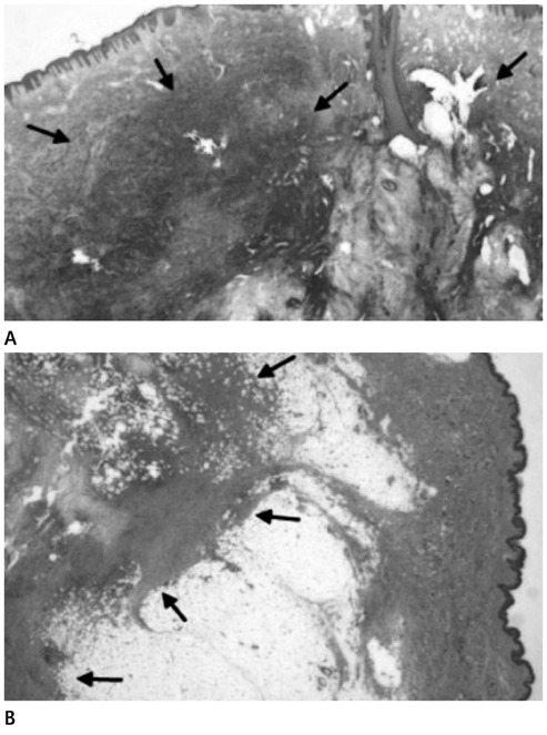
Fig. 6 The final pathologic results confirmed metastatic dermato fibrosarcoma protuberans with fibrosarcomatous changes. A. Pathologic specimen of an initial abdominal lesion showed sarcomatous changes (arrows) in dermatofibrosarcoma protuberans arising in the dermis (hematoxylin-eosin, × 12). B. Pathologic specimen of a recurrent axillary lesion showed similar histologic freatures of sarcomatous changes in dermatofibrosarcoma protuberans (arrows) locating in subcutaneous fatty layer (hematoxylin-eosin, × 12).
Reference
-
1. Bogucki B, Neuhaus I, Hurst EA. Dermatofibrosarcoma Protuberans: A Review of the Literature. Dermatol Surg. 2012. [Epub ahead of print].2. Bowne WB, Antonescu CR, Leung DH, Katz SC, Hawkins WG, Woodruff JM, et al. Dermatofibrosarcoma protuberans: a clinicopathologic analysis of patients treated and followed at a single institution. Cancer. 2000. 88:2711–2720.3. Erdem O, Wyatt AJ, Lin E, Wang X, Prieto VG. Dermatofibrosarcoma protuberans treated with wide local excision and followed at a cancer hospital: prognostic significance of clinicopathologic variables. Am J Dermatopathol. 2012. 34:24–34.4. Mendenhall WM, Zlotecki RA, Scarborough MT. Dermatofibrosarcoma protuberans. Cancer. 2004. 101:2503–2508.5. Abbott JJ, Oliveira AM, Nascimento AG. The prognostic significance of fibrosarcomatous transformation in dermatofibrosarcoma protuberans. Am J Surg Pathol. 2006. 30:436–443.6. Ding J, Hashimoto H, Enjoji M. Dermatofibrosarcoma protuberans with fibrosarcomatous areas. A clinicopathologic study of nine cases and a comparison with allied tumors. Cancer. 1989. 64:721–729.7. Mentzel T, Beham A, Katenkamp D, Dei Tos AP, Fletcher CD. Fibrosarcomatous ("high-grade") dermatofibrosarcoma protuberans: clinicopathologic and immunohistochemical study of a series of 41 cases with emphasis on prognostic significance. Am J Surg Pathol. 1998. 22:576–587.8. Palmerini E, Gambarotti M, Staals EL, Zanella L, Sieberova G, Longhi A, et al. Fibrosarcomatous changes and expression of CD34+ and apolipoprotein-D in dermatofibrosarcoma protuberans. Clin Sarcoma Res. 2012. 2:4.9. Goldblum JR, Reith JD, Weiss SW. Sarcomas arising in dermatofibrosarcoma protuberans: a reappraisal of biologic behavior in eighteen cases treated by wide local excision with extended clinical follow up. Am J Surg Pathol. 2000. 24:1125–1130.10. Kransdorf MJ, Meis-Kindblom JM. Dermatofibrosarcoma protuberans: radiologic appearance. AJR Am J Roentgenol. 1994. 163:391–394.11. Lee SJ, Mahoney MC, Shaughnessy E. Dermatofibrosarcoma protuberans of the breast: imaging features and re view of the literature. AJR Am J Roentgenol. 2009. 193:W64–W69.12. Shin YR, Kim JY, Sung MS, Jung JH. Sonographic findings of dermatofibrosarcoma protuberans with pathologic correlation. J Ultrasound Med. 2008. 27:269–274.13. Torreggiani WC, Al-Ismail K, Munk PL, Nicolaou S, O'Connell JX, Knowling MA. Dermatofibrosarcoma protuberans: MR imaging features. AJR Am J Roentgenol. 2002. 178:989–993.
