Severe Endobronchial Inflammation Induced by Aspiration of a Ferrous Sulfate Tablet
- Affiliations
-
- 1Division of Respiratory and Critical Care Medicine, Department of Internal Medicine, Korea University Guro Hospital, Korea University Medical School, Seoul, Korea. minkyunghoon@korea.ac.kr
- KMID: 2320754
- DOI: http://doi.org/10.4046/trd.2016.79.1.37
Abstract
- Iron supplements such as ferrous sulfate tablets are usually used to treat iron-deficiency anemia in some elderly patients with primary neurologic disorders or decreased gag reflexes due to stroke, senile dementia, or parkinsonism. While the aspiration of ferrous sulfate is rarely reported, it is a potentially life-threatening condition that can lead to airway necrosis and bronchial stenosis. A detailed history and high suspicion of aspiration are required to avoid delays in diagnosis and treatment. The diagnosis can be confirmed by bronchoscopic examination and a tissue biopsy. Early removal of the aspirated tablet prevents acute complications, such as bronchial necrosis, hemoptysis, and lobar consolidation. Tablet removal is also necessary to prevent late bronchial stenosis. We presented the first case in Korea of a ferrous sulfate tablet aspiration that induced severe endobronchial inflammation.
Keyword
MeSH Terms
Figure
-

Figure 1 (A, B) The admission chest X-ray showed atelectasis of the right middle lung and consolidations of the right lower lung, with thickening of the right minor fissure: posteroanterior (A) and lateral views (B). (C-F) Chest computed tomography on hospital day 5 showed a high-density lesion in the right bronchus intermedius, atelectasis in right middle and lower lobes, consolidations and ground-glass opacities in the right upper and lower lobes, and a right pleural effusion: mediastinal (C, E) and lung (D, F) settings. (G, H) Chest X-ray 1 month after the end of treatment showed near-complete resolution of the lung lesions seen on admission.
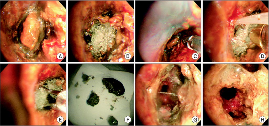
Figure 2 (A, B) The bronchoscopic examination showed near-total obstruction of the right bronchus intermedius due to impacted pill-like material, which appeared greyish brown and fragile. The endobronchial mucosa around this material showed severe edematous inflammatory and friable changes, with a touch bleeding and yellowish brown discoloration. (C-G) Procedure images showing removal of the pill-like material with grasp biopsy forceps, W-shaped grasping forceps, and a cryotherapy system with grasp biopsy forceps (C), with the cryotherapy system (D), with W-shaped grasping forceps (E), removed pill-like materials (F), and right bronchus intermedius after removal (G). (H) Bronchoscopic examination 1 week after removing the tablet showed that the previous severe endobronchial inflammation of the right bronchus intermedius was improved, but still remained.

Figure 3 (A-D) The pathological results of the removed pill-like material and biopsy tissue around the pill-like material in the right bronchus intermedius revealed aspiration of ferrous sulfate. (A) Removed pill-like material. (B) Bronchus intermedius (A and B, H&E stain, ×100; C, H&E stain, ×400; D, iron stain, ×400).
Reference
-
1. Baharloo F, Veyckemans F, Francis C, Biettlot MP, Rodenstein DO. Tracheobronchial foreign bodies: presentation and management in children and adults. Chest. 1999; 115:1357–1362.2. Lamaze R, Trechot P, Martinet Y. Bronchial necrosis and granuloma induced by the aspiration of a tablet of ferrous sulphate. Eur Respir J. 1994; 7:1710–1711.3. Boyd M, Watkins F, Singh S, Haponik E, Chatterjee A, Conforti J, et al. Prevalence of flexible bronchoscopic removal of foreign bodies in the advanced elderly. Age Ageing. 2009; 38:396–400.4. Lee JO, Lee JH, Ahn S, Kim JW, Chang H, Kim YJ, et al. Prevalence and risk factors for iron deficiency anemia in the korean population: results of the fifth KoreaNational Health and Nutrition Examination Survey. J Korean Med Sci. 2014; 29:224–229.5. Mahmood K, Koubar S, Shofer SL, Ninan NA, Wahidi MM. Alendronate tracheobronchitis. Ann Am Thorac Soc. 2013; 10:64–66.6. Sundar KM, Elliott CG, Thomsen GE. Tetracycline aspiration: case report and review of the literature. Respiration. 2001; 68:416–419.7. Karakan Y, Akpinar A, Yildiz H, Aksoy H, Dikensoy O. A case of ciprofloxacin tablet aspiration. Tuberk Toraks. 2010; 58:97–99.8. Micallef J, Montefort S, Mallia Azzopardi C, Galea J. Two cases of aspiration of calcium tablets. Lung India. 2011; 28:312–314.9. Radiological notes: aspirated capsule in right lower lobe. J Mt Sinai Hosp N Y. 1966; 33:530–532.10. Cimino-Mathews A, Illei PB. Cytologic and histologic findings of iron pill-induced injury of the lower respiratory tract. Diagn Cytopathol. 2013; 41:901–903.11. Maw M, Chiu R, Lim AY. Bronchoscopic and histological changes over time following acute ferrous sulphate tablet aspiration. BMJ Case Rep. 2012; 2012:bcr2012007329.12. Venci NM, Watson TJ, Kallay MC. Bronchial stenosis following ferrous sulfate aspiration: case report and review of the literature. J Bronchology Interv Pulmonol. 2014; 21:58–60.13. Ceylan N, Bayraktaroglu S, Savas R, Alper H. CT findings of high-attenuation pulmonary abnormalities. Insights Imaging. 2010; 1:287–292.14. Lee P, Culver DA, Farver C, Mehta AC. Syndrome of iron pill aspiration. Chest. 2002; 121:1355–1357.15. Kim ST, Kaisar OM, Clarke BE, Vandenburg RA, Allen DH, Bell SC, et al. 'Iron lung': distinctive bronchoscopic features of acute iron tablet aspiration. Respirology. 2003; 8:541–543.
- Full Text Links
-
- Actions
-
Cited
- CITED
-
- Close
- Share
-
- Similar articles
-
- A Case of Significant Endobronchial Injury due to Recurrent Iron Pill Aspiration
- Iron Supplementation in a Girl with Attention-Deficit Hyperactivity Disorder
- The Effect of Splenectomy on Acute Barium Sulfate Induced Toxicity of Rat Organs
- Hyponatremic Seizure after Ingestion of an Oral Sulfate Tablet for Bowel Preparation for Colonoscopy
- Effects of Iron Supplementation on Attention Deficit Hyperactivity Disorder in Children Treated with Methylphenidate
