Endobronchial ALK-Positive Anaplastic Large Cell Lymphoma Presenting Massive Hemoptysis
- Affiliations
-
- 1Department of Internal Medicine, CHA Bundang Medical Center, CHA University School of Medicine, Seongnam, Korea. jhcmd@cha.ac.kr
- 2Department of Pathology, CHA Bundang Medical Center, CHA University School of Medicine, Seongnam, Korea.
- 3Divison of Respiratory and Critical Care Medicine, Department of Internal Medicine, CHA Bundang Medical Center, CHA University School of Medicine, Seongnam, Korea.
- KMID: 2320713
- DOI: http://doi.org/10.4046/trd.2015.78.4.390
Abstract
- Primary anaplastic large cell lymphoma (ALCL) of the lung is highly aggressive and quite rare. We report here a case of anaplastic lymphoma kinase-positive endobronchial ALCL, that was initially thought to be primary lung cancer. A 68-year-old woman presented with hemoptysis, dyspnea, and upper respiratory symptoms persisting since 1 month. The hemoptysis and and bronchial obstruction lead to respiratory failure, prompting emergency radiotherapy and steroid treatment based on the probable diagnosis of lung cancer, although a biopsy did not confirm malignancy. Following treatment, her symptoms resolved completely. Chest computed tomography scan performed 8 months later showed increased and enlarged intra-abdominal lymph nodes, suggesting lymphoma. At that time, a lymph node biopsy was recommended, but the patient refused and was lost to follow up. Sixteen months later, the patient revisited the emergency department, complaining of persistent abdominal pain since several months. A laparoscopic intra-abdominal lymph node biopsy confirmed a diagnosis of ALCL.
Keyword
MeSH Terms
Figure
-
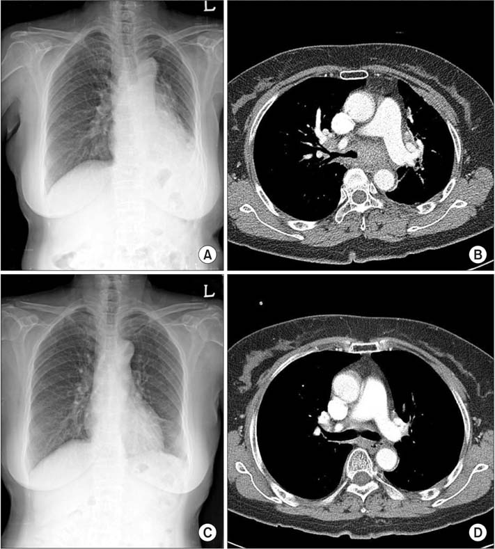
Figure 1 An initial chest X-ray (A) and chest computed tomography (B) revealed obstruction of the left main bronchus with obstructive collapse with air trapping in the left lung, which suggested lung cancer with obstructive collapse. Chest X-ray (C) and chest CT (D), taken on the 12th day after radiotherapy and steroid administration, revealed a markedly decreased mass and disappearance of obstruction in the left main bronchus.
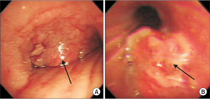
Figure 2 Bronchoscopy showed total obstruction of the left main bronchus (A, arrow indicates the mass located at proximal left main bronchus extending carina). Bronchoscopy done after radiotherapy showed no evidence of bleeding and resolved obstruction (B, arrow indicates the remnant of mass).
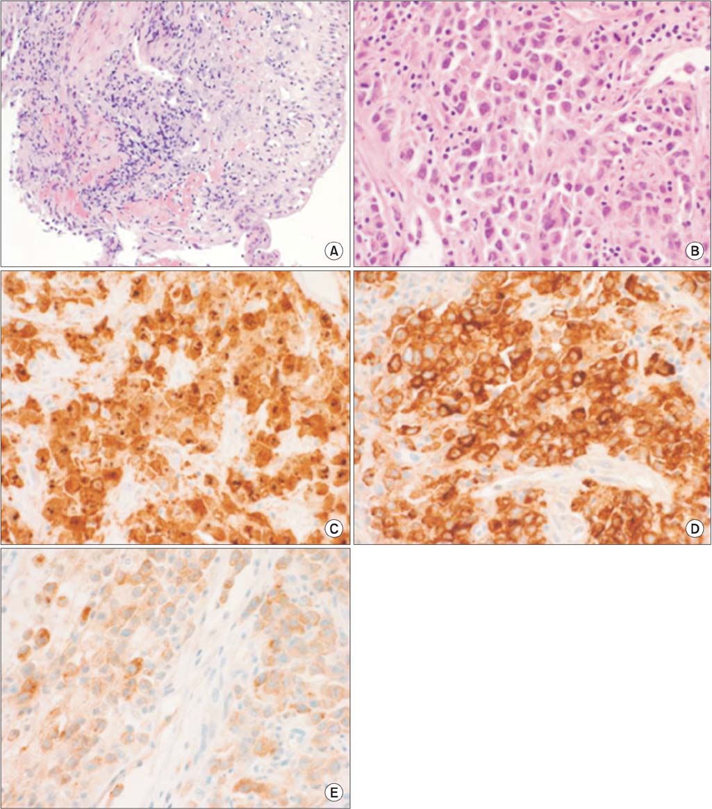
Figure 3 The biopsy performed on her first hospitalization showed only ulceration and granulation tissue (A, H&E stain, ×100). Laparoscopic biopsy from intra-abdominal lymph nodes shows anaplastic large cell lymphoma cells having marked pleomorphism (B, H&E stain, ×400). Immunohistochemical stain reveals positivity for anaplastic lymphoma kinase (C, ×400), epithelial membrane angiten (D, ×400), and CD30 (E, ×400).
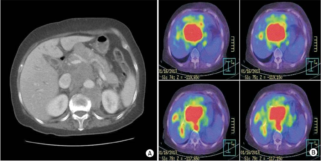
Figure 4 Abdomen-pelvis computed tomography (A) revealed extensive intra-abdominal lymph node metastasis and biliary obstruction by markedly increased size of portocaval and hepatoduodenal ligament, which showed as hypermetabolic lesions in positron emission tomography (B).
Reference
-
1. Medeiros LJ, Elenitoba-Johnson KS. Anaplastic large cell lymphoma. Am J Clin Pathol. 2007; 127:707–722.2. Kim JH, Lee SH, Park J, Kim HY, Lee SI, Park JO, et al. Primary pulmonary non-Hodgkin's lymphoma. Jpn J Clin Oncol. 2004; 34:510–514.3. Rush WL, Andriko JA, Taubenberger JK, Nelson AM, Abbondanzo SL, Travis WD, et al. Primary anaplastic large cell lymphoma of the lung: a clinicopathologic study of five patients. Mod Pathol. 2000; 13:1285–1292.4. Barthwal MS, Deoskar RB, Falleiro JJ, Singh P. Endobronchial non-Hodgkin's lymphoma. Indian J Chest Dis Allied Sci. 2005; 47:117–120.5. Xu X. ALK-negative anaplastic large cell lymphoma primarily involving the bronchus: a case report and literature review. Int J Clin Exp Pathol. 2014; 7:460–463.6. Han SH, Maeng YH, Kim YS, Jo JM, Kwon JM, Kim WK, et al. Primary anaplastic large cell lymphoma of the lung presenting with acute atelectasis. Thorac Cancer. 2014; 5:78–81.7. Ferraro P, Trastek VF, Adlakha H, Deschamps C, Allen MS, Pairolero PC. Primary non-Hodgkin's lymphoma of the lung. Ann Thorac Surg. 2000; 69:993–997.8. Yang HB, Li J, Shen T. Primary anaplastic large cell lymphoma of the lung. Report of two cases and literature review. Acta Haematol. 2007; 118:188–191.9. Lee SA, Kim DH, Jeon GS. Covered bronchial stent insertion to manage airway obstruction with hemoptysis caused by lung cancer. Korean J Radiol. 2012; 13:515–520.
- Full Text Links
-
- Actions
-
Cited
- CITED
-
- Close
- Share
-
- Similar articles
-
- CD30-Positive Anaplastic Lymphoma Kinase-Negative Systemic Anaplastic Large-Cell Lymphoma in a 9-Year-Old Boy
- A Case of ALK-Negative Systemic Anaplastic Large Cell Lymphoma
- A Case of Multiple Cranial Neuropathies Caused by Anaplastic Lymphoma Kinase-Negative Anaplastic Large Cell Lymphoma
- A Case of CD 30+/ALK− Primary Systemic Anaplastic Large Cell Lymphoma Presenting a Sporotrichoid Pattern
- A Case of CD30 (+)/ALK (-) Systemic Anaplastic Large Cell Lymphoma
