Tuberc Respir Dis.
2011 Nov;71(5):322-327.
Value of Bronchoalveolar Lavage Fluid Cytology in the Diagnosis of Pneumocystis jirovecii Pneumonia: A Review of 30 Cases
- Affiliations
-
- 1Department of Pathology, Samsung Medical Center, Sungkyunkwan University School of Medicine, Seoul, Korea. hanjho@skku.edu
- 2Division of Pulmonary and Critical Care Medicine, Department of Medicine, Samsung Medical Center, Sungkyunkwan University School of Medicine, Seoul, Korea.
- 3Department of Pathology, Gachon University Gil Hospital, Incheon, Korea.
Abstract
- BACKGROUND
Pneumocystis jirovecii is a fungus that has become an important cause of opportunistic infections. We present a summary of the clinical status and findings from bronchoalveolar lavage (BAL) of patients with Pneumocystis jirovecii pneumonia (PJP).
METHODS
We selected 30 cases of PJP that were proven through a surgical specimen evaluation. BAL fluid cytology was reviewed, and agreement with the initial diagnosis was evaluated.
RESULTS
All 30 cases of PJP occurred in immunocompromised patients. Only 15 of the 30 cases were initially diagnosed as PJP. We found PJP in 13 of the 15 cases that were negative at the initial diagnosis. The most characteristic finding of PJP was frothy exudates, and BAL fluid tended to show rare neutrophils. Two of seven patients with PJP and diffuse alveolar damage (DAD) revealed no frothy exudates in BAL fluid.
CONCLUSION
BAL fluid cytology was reconfirmed as a sensitive and rapid method to diagnose PJP. We must be aware of the possibility of PJP to maintain high diagnostic sensitivity. We cannot exclude PJP in cases of PJP with DAD, even if frothy exudates are not observed in the BAL fluid.
MeSH Terms
Figure
-
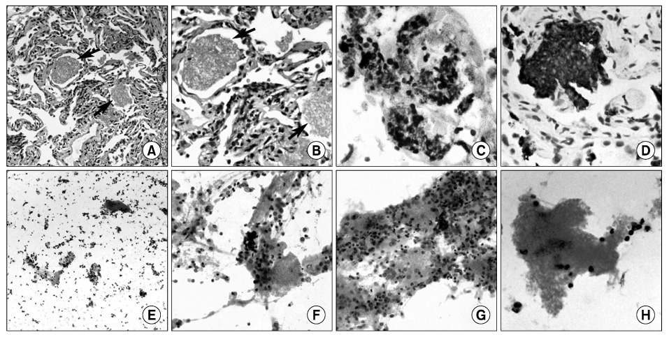
Figure 1 Characteristic PJP findings in surgical specimens (A~D) and bronchoalveolar fluid (E~H). (A, B) Frothy, foamy, or honeycomb exudates (arrow) within the alveolar spaces (A, H&E, ×100; B, H&E, ×200). (C, D) GMS staining and immunohistochemistry for Pneumocystis highlights PJP organisms (C, GMS, ×400; D, immunohistochemistry, ×400). (E~H) Scattered frothy exudates intermixed with inflammatory cells (E, H&E, ×40; F, H&E, ×100; H, H&E, ×400). PJP: Pneumocystis jirovecii pneumonia; GMS: gomori methenamine-silver.
Reference
-
1. Sidhu GS, Cassai ND, Pei Z. Pneumocystis carinii: an update. Ultrastruct Pathol. 2003. 27:115–122.2. Haque AK. Zander DS, Farver CF, editors. Fungal disease. Pulmonary pathology. 2008. 1st ed. Philadelphia: Churchill Livingstone;236–239.3. Willebrand Ev, Lautenschlager I. Gray W, Kocjan G, editors. Organ transplantation. Diagnostic cytopathology. 2010. 3rd ed. Edinburgh: Chrchill Livingstone/Elsevier;475–476.4. Jones JL, Hanson DL, Dworkin MS, Alderton DL, Fleming PL, Kaplan JE, et al. Surveillance for AIDS-defining opportunistic illnesses, 1992-1997. MMWR CDC Surveill Summ. 1999. 48:1–22.5. Choe KW, Oh MD, Park SW, Kim HB, Kim US, Kang SW, et al. Opportunistic infections and malignancies in 173 patients with HIV infection. Korean J Infect Dis. 1998. 30:507–515.6. Kim JM, Cho GJ, Hong SK, Chang KH, Chung JS, Choi YH, et al. Epidemiology and clinical features of HIV infection/AIDS in Korea. Yonsei Med J. 2003. 44:363–370.7. Greaves TS, Strigle SM. The recognition of Pneumocystis carinii in routine Papanicolaou-stained smears. Acta Cytol. 1985. 29:714–720.8. Price RA, Hughes WT. Histopathology of Pneumocystis carinii infestation and infection in malignant disease in childhood. Hum Pathol. 1974. 5:737–752.9. Broaddus C, Dake MD, Stulbarg MS, Blumenfeld W, Hadley WK, Golden JA, et al. Bronchoalveolar lavage and transbronchial biopsy for the diagnosis of pulmonary infections in the acquired immunodeficiency syndrome. Ann Intern Med. 1985. 102:747–752.10. Golden JA, Hollander H, Stulbarg MS, Gamsu G. Bronchoalveolar lavage as the exclusive diagnostic modality for Pneumocystis carinii pneumonia. A prospective study among patients with acquired immunodeficiency syndrome. Chest. 1986. 90:18–22.11. Orenstein M, Webber CA, Cash M, Heurich AE. Value of bronchoalveolar lavage in the diagnosis of pulmonary infection in acquired immune deficiency syndrome. Thorax. 1986. 41:345–349.12. Chechani V, Allam AA, Haseeb MA, Kamholz SL. Pneumocystis carinii pneumonia in patients with AIDS: evaluation of lavage and staining techniques in diagnosis. J Acquir Immune Defic Syndr. 1991. 4:250–253.13. Lee JH, Lee JY, Shin MR, Ahn HK, Kim CW, Kim I. Immunohistochemical identification of pneumocystis jirovecii in liquid-based cytology of bronchoalveolar lavage: nine cases report. Korean J Pathol. 2011. 45:115–118.14. Ziefer A, Abramowitz JA. Pneumocystis carinii pneumonia in HIV-positive and HIV-negative patients. An epidemiological, clinical and histopathological study of 18 patients. S Afr Med J. 1989. 76:308–313.15. Burke BA, Good RA. Pneumocystis carinii infection. Medicine (Baltimore). 1973. 52:23–51.
- Full Text Links
-
- Actions
-
Cited
- CITED
-
- Close
- Share
-
- Similar articles
-
- Correspondence Regarding the Article Titled “Low Lymphocyte Proportion in Bronchoalveolar Lavage Fluid as a Risk Factor Associated with the Change from Trimethoprim/sulfamethoxazole Used as First-Line Treatment for Pneumocystis jirovecii Pneumonia”
- Reply: Correspondence Regarding the Article Titled “Low Lymphocyte Proportion in Bronchoalveolar Lavage Fluid as a Risk Factor Associated with the Change from Trimethoprim/sulfamethoxazole Used as First-Line Treatment for Pneumocystis jirovecii Pneumonia”
- Immunohistochemical Identification of Pneumocystis jirovecii in Liquid-based Cytology of Bronchoalveolar Lavage: Nine Cases Report
- Early diagnosis of pneumocystis carinii pneumonia by calcofluor white stain in bronchoalveolar lavage fluid
- Spontaneous Pneumomediastinum, Pneumopericardium, and Pneumothorax with Respiratory Failure in a Patient with AIDS and Pneumocystis jirovecii Pneumonia
