2 Cases of a Benign Pulmonary Metastasizing Leiomyoma
- Affiliations
-
- 1Division of Respiratory and Critical Care Medicine, Department of Internal Medicine, Korea University Hospital, Seoul, Korea. khin@kumc.or.kr
- 2Department of Obstetrics and Gynecology, Korea University Hospital, Seoul, Korea.
- 3Department of Pathology, Korea University Hospital, Seoul, Korea.
- 4Department of Radiology, Korea University Hospital, Seoul, Korea.
Abstract
- A benign pulmonary metastasizing leiomyoma is a recognized clinical entity that has been infrequently reported in the medical literature. We report two cases of a benign pulmonary metastasizing leiomyoma. A 35-year-old woman who underwent myomectomy and a cesarean section approximately 6 years earlier visited our hospital for further evaluation of incidentally revealed multiple lung nodules. A diagnostic percutaneuous biopsy was performed. Finally she was diagnosed with a benign metastasizing leiomyoma. The patient then received LH-RH and has been followed up since. The other 44-year-old woman presented after an initial radiology evaluation revealed the presence of multiple, small-sized lung nodules. She underwent a right middle lung wedge resection to confirm the diagnosis. Finally she diagnosed with a benign metastasizing leiomyoma. The multiple lung nodules have been followed up closely.
MeSH Terms
Figure
-
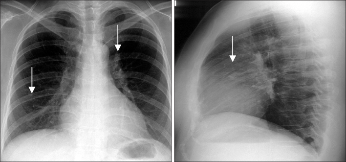
Figure 1 Chest radiograph shows multiple lung nodules.
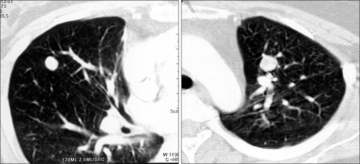
Figure 2 Chest CT shows two round, well-defined and poorly enhanced nodules.
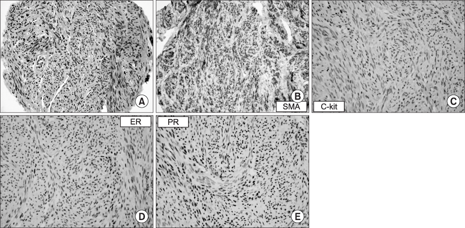
Figure 3 (A) Microscopic examination of a percutaneous needle biopsy specimen of the lung nodule shows intersecting bundles of spindle-shaped cells, which are strongly-positive for (B) smooth muscle actin immunohistochemical stain, and they highly express both (D) estrogen and (E) progesterone. However, the tumor cells are negative for (C) C-kit (CD 117) immunohistochemical stain (A, H&E stain, ×400).
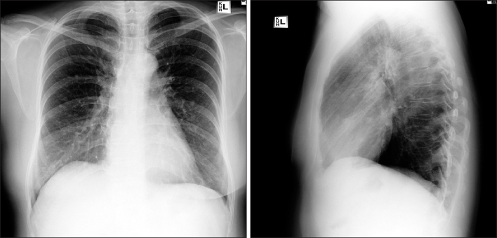
Figure 4 Chest radiograph shows multiple, round lung nodules.
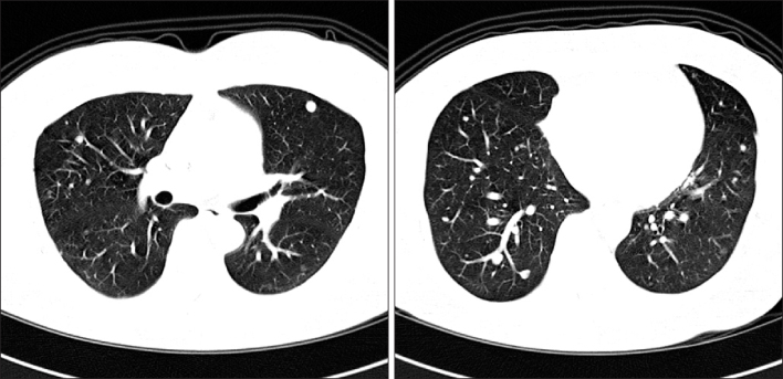
Figure 5 Chest CT reveals multiple nodules ranging in diameter from a few millimeters to 2 cm.

Figure 6 (A) The resected nodule is clearly demarcated from thesurrounding lung tissue. (B) It shows thetypical features of a leiomyoma with no findings suggestive of malignant potential such as coagulative necrosis, increased mitotic activity or significant atypia. (C) The low cuboidal metaplastic bronchiolar epithelia are invaginated and entrapped between the fascicles of the smooth muscle (H&E stain, A, ×40, B, ×200, C, ×200).
Reference
-
1. Giove S, Scappaticci E, Baldi S, Ricci C, Minetto E. Benign metastasizing leiomyoma of the uterus: case report. Minerva Med. 1984. 75:1819–1821.2. Abramson S, Gilkeson RC. Multiple pulmonary nodules in an asymptomatic patient. Chest. 1999. 116:245–247.3. Abramson S, Gilkeson RC, Goldstein JD, Woodard PK, Eisenberg R, Abramson N. Benign metastasizing leiomyoma: clinical, imaging, and pathologic correlation. AJR Am J Roentogenol. 2001. 176:1409–1413.4. Esteban JM, Allen WM, Schaerf RH. Benign metastasizing leiomyoma of the uterus: histologic and immunohistochemical characterization of primary and metastatic lesions. Arch Pathol Lab Med. 1999. 123:960–962.5. Houck WV, Broderick TJ, Cohen SA, Cohen NM. Benign metastasizing leiomyoma. Surg Endosc. 2002. 16:716.6. Tietze L, Gunther K, Horbe A, Pawlik C, Klosterhalfen B, Handt S, et al. Benign metastasizing leiomyoma: a cytogenetically balanced but clonal disease. Hum Pathol. 2000. 31:126–128.7. Arai T, Yasuda Y, Takaya T, Shibayama M. Natural decrease of benign metastasizing leiomyoma. Chest. 2000. 117:921–922.8. Kang SA, Choi SI, Kim YA, Kim CJ, Yang DG, Kang JH, et al. A case of benign metastasizing pulmonary leimyoma. Tuberc Respir Dis. 2005. 58:614–618.9. Hwang JK, Park KY, Park JW, Park JK, Jeong SH, Jeong JB, et al. A case of benign metastasizing leiomyoma in the lung. Tuberc Respir Dis. 2000. 49:231–236.
- Full Text Links
-
- Actions
-
Cited
- CITED
-
- Close
- Share
-
- Similar articles
-
- A Case of Benign Metastasizing Leiomyoma of Uterus to the Lung
- A Case of Benign Metastasizing Pulmonary Leiomyomatosis
- Natural Course of Benign Pulmonary Metastasizing Leiomyoma
- Incidental Multiple Pulmonary Nodules: Benign Metastasizing Leiomyoma and 18F-FDG PET/CT
- A Case of Metastasis to Lung of Benign Metastasizing Leiomyoma
