A Case of Growing Endobronchial Glandular Papilloma
- Affiliations
-
- 1Division of Pulmonary and Critical Care Medicine, Department of Internal Medicine, Pusan Paik Hospital, Inje University School of Medicine, Busan, Korea. goodoc@gmail.com
- 2Department of Pathology, Pusan Paik Hospital, Inje University School of Medicine, Busan, Korea.
- 3Department of Radiology, Pusan Paik Hospital, Inje University School of Medicine, Busan, Korea.
- 4Department of Rehabilitation, Pusan Paik Hospital, Inje University School of Medicine, Busan, Korea.
Abstract
- Pulmonary papillomas are rare benign epithelial neoplasms arising in bronchial surface epithelium. They are categorized by a variety of cell types including squamous, glandular, and mixed squamous and glandular type. Among them, glandular papilloma is extremely rare and has not been reported in Korea. The patient was a 52 year-old man presenting with a 4-months' history of recurrent hemoptysis. Bronchofiberoscopy revealed a whitish, glistening, and polypoid mass lesion at the proximal bronchus in the basal segment of the left lower lung. Bronchoscopic biopsy was performed; papillary fronds lined by ciliated or nonciliated pseudostratified columnar epithelium were noted on histologic findings. We present the first case of glandular papilloma in Korea. Two years later, the patient visited our hospital again due to hemoptysis. On follow-up bronchoscopy, a mass that had been found previously showed an increase in size.
Keyword
MeSH Terms
Figure
-
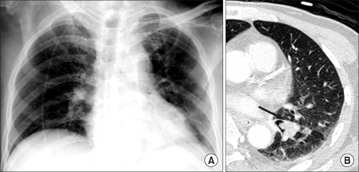
Figure 1 (A) Supine anteriorposterior chest radiograph shows mild volume loss and patch consolidation in the left lower lobe. (B) Axial CT scan show endobronchial mass (arrow) in the proximal portion of left lower basal segmental bronchus and atelectasis in the medial and anterior basal segment of left lower lobe.

Figure 2 (A) Initial bronchoscopy shows a glistening, polypoid and lobulating mass lesion that partially obstructing the orifice of left lower lobe basal segment. (B) Follow up bronchoscopy shows a increased mass lesion that completely occluding the lumen of left lower lobe bronchus.

Figure 3 Endobrochial biopsied tissue reveals papillary fronds lined by pseudostratified nonciliated or focally ciliated columnar cells showing piliform cellular tufting (H&E stain, ×100).
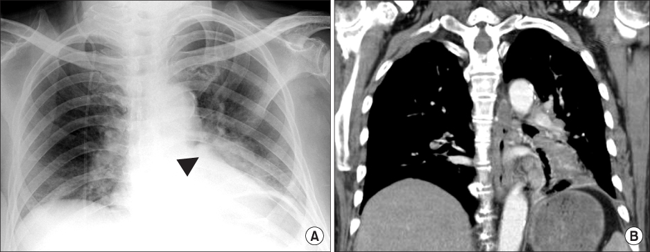
Figure 4 (A, B) Follow up supine anteriorposterior chest radiograph and coronal CT scan after 2 years show more enlarged endobronchial mass (arrowhead) and progression of postobstructive atelectasis and consolidation in the basal segment of left lower lobe.
Reference
-
1. Travis WD, Brambilla E, Muller-Hermelink HK, Harris CC. World Health Organization. Travis , editor. Pathology and genetics of tumors of the lung, pleura, thymus and heart. 2004. Lyon: IARC Press;78–81.2. Ashmore PG. Papilloma of the bronchus: case report. J Thorac Surg. 1954. 27:293–294.3. Flieder DB, Koss MN, Nicholson A, Sesterhenn IA, Petras RE, Travis WD. Solitary pulmonary papillomas in adults: a clinicopathologic and in situ hybridization study of 14 cases combined with 27 cases in the literature. Am J Surg Pathol. 1998. 22:1328–1342.4. Basheda S, Gephardt GN, Stoller JK. Columnar papilloma of the bronchus: case report and literature review. Am Rev Respir Dis. 1991. 144:1400–1402.5. Sung CO, Kim J, Do IG, Han J. Solitary pulmonary mixed squamous cell and glandular papilloma: a brief case report. Korean J Pathol. 2008. 42:393–395.6. Popper HH, Wirnsberger G, Jüttner-Smolle FM, Pongratz MG, Sommersgutter M. The predictive value of human papilloma virus (HPV) typing in the prognosis of bronchial squamous cell papillomas. Histopathology. 1992. 21:323–330.7. Spencer H, Dail DH, Arneaud J. Non-invasive bronchial epithelial papillary tumors. Cancer. 1980. 45:1486–1497.8. Kwon JE, Kim GY, Han J, Kim TS, Kim K. Mucous gland adenoma presenting as a peripheral lung mass: a brief case report. Korean J Pathol. 2004. 38:126–128.9. Sohn ST, Jeong TY, Lee WM, Kang JH, Kim H, Chung WS, et al. Papillary adenoma of the lung with pulmonary. sequestration: a case report. Korean J Thorac Cardiovasc Surg. 1997. 30:1262–1266.10. Ashley DJ, Danino EA, Davies HD. Bronchial polyps. Thorax. 1963. 18:45–49.
- Full Text Links
-
- Actions
-
Cited
- CITED
-
- Close
- Share
-
- Similar articles
-
- Glandular papilloma of the lung with malignant transformation
- Mixed Squamous Cell and Glandular Papilloma of the Lung in a 64-Year-Old Woman
- Solitary Pulmonary Mixed Squamous Cell and Glandular Papilloma: A Brief Case Report
- Mixed Squamous Cell and Glandular Papilloma Presented with Peripheral Lung Mass: A Case Report
- A Case of Bronchial Papilloma
