2 Cases of Mycoplasma pneumoniae Infection with Severe Pneumonia
- Affiliations
-
- 1Department of Internal Medicine, Yonsei University Wonju College of Medicine, Wonju, Korea. sjyong@yonsei.ac.kr
Abstract
- Mycoplasma pneumoniae (M. pneumoniae) is the leading cause of pneumonia in older children and young adults. Normally, it does not progress to a condition requiring hospitalization but improves spontaneously or has a mild clinical course. We report two cases of M. pneumoniae pneumonia with different clinical manifestations from the normal course. The patients were young healthy individuals. The diagnoses were made by serology. However, it could not be determined beforehand that they had M. pneumoniae pneumonia. Based on the empirical treatment strategy of severe community acquired pneumonia, the patients were treated with broad-spectrum antibiotics including cephalosporin, quinolone and macrolide. After administering the antibiotics, they showed a gradually favorable clinical course and recovered without residual complications. A M. pneumoniae infection should be considered as a cause of severe community acquired pneumonia, and empirical treatment targeting this organism might be helpful in treating patients with the severe manifestation.
Keyword
MeSH Terms
Figure
-
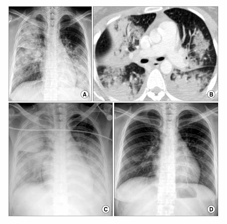
Figure 1 A 29-year-old woman with M. pneumoniae pneumonia. Chest radiograph at admission reveals bilateral, patch and lobar consolidation in right upper and left lower lung field (A). Chest CT scan, 2 days later, shows muti-focal consolidation (B). Follow up chest radiograph, 3days later, shows more extensive bilateral consolidation (C). Follow up chest radiograph, 9 days later, shows nearly complete resolution (D).
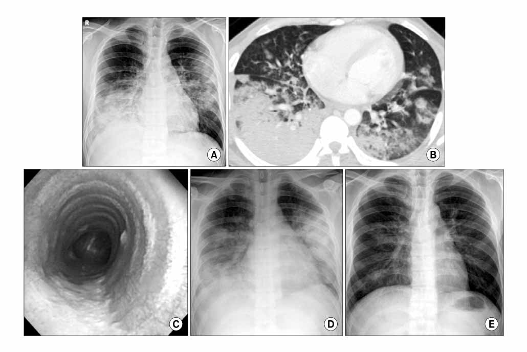
Figure 2 A 29-year-old man with M. pneumoniae pneumonia. Chest radiograph at admission reveals bilateral, patch and lobar consolidation in right lower and left upper lung field (A). Chest CT scan shows muti-focal lobar consolidation, mainly in right lower lobe, with pleural effusion (B). Fiberoptic bronchoscopy shows ulcerative lesion covered with necrotic exudation on the posterior wall of trachea (C). Follow up chest radiograph, 3days later, shows more aggravated consolidation (D). Follow up chest radiograph, 13 days later, shows improved state (E).
Reference
-
1. Cassell GH. Severe Mycoplasma disease: rare or underdiagnosed? West J Med. 1995. 162:172–175.2. Murray JF, Nadel JA. Textbook of respiratory medicine. 2005. 4th ed. Philadelphia: Elsevier Saunders.3. Petitjean J, Vabret A, Gouarin S, Freymuth F. Evaluation of four commercial immunoglobulin G (IgG)- and IgM-specific enzyme immunoassays for diagnosis of Mycoplasma pneumoniae infections. J Clin Microbiol. 2002. 40:165–171.4. Jang BI, Kim HI, Kim SS, Lee CK, Chung JH, Lee KH, et al. A case of Mycoplasma pneumonia complicated with acute respiratory failure. Tuberc Respir Dis. 1992. 39:194–198.5. Kim ES, Lee WS, Lee KR, Lee JA, Baek YJ, Lee GS, et al. A case of Mycoplasma pneumonia which progressed to ARDS. Tuberc Respir Dis. 1996. 43:645–650.6. Kwag JS, Ko TY, Chung BS, Lee SB, Oh KS, Park SJ, et al. A case of fulminant Mycoplasma pneumonia complicated with ARDS and hemolytic anemia. Tuberc Respir Dis. 1998. 46:636–642.7. Choi SM, Han CM, Kang JH, Chang WI, Kim CH, Kim KH, et al. Extensive bilateral airspace consolidation. Tuberc Respir Dis. 1999. 46:735–740.8. Yu CW, Cheong HJ, Kang MS, Woo HJ, Kim WJ, Kim MJ, et al. A case Mycoplasma pneumoniae pneumonia with acute respiratory distress syndrome (ARDS). Korean J Med. 2000. 70:94–100.9. Chan ED, Welsh CH. Fulminant Mycoplasma pneumoniae pneumonia. West J Med. 1995. 162:133–142.10. Fraley DS, Ruben FL, Donnelly EJ. Respiratory failure secondary to Mycoplasma pneumoniae infection. South Med J. 1979. 72:437–440.11. Taylor-Robinson D, Furr PM. Models of infection due to mycoplasmas, including Mycoplasma fermentans, in the genital tract and other sites in mice. Clin Infect Dis. 1993. 17:S280–S282.12. Brunner H, Horswood RL, Chanock RM. More sensitive methods for detection of antibody to Mycoplasma pneumoniae. J Infect Dis. 1973. 127:S52–S55.13. Waites KB, Talkington DF. Mycoplasma pneumoniae and its role as a human pathogen. Clin Microbiol Rev. 2004. 17:697–728.
- Full Text Links
-
- Actions
-
Cited
- CITED
-
- Close
- Share
-
- Similar articles
-
- A Case of Cerebral Infarction Complicated by Mycoplasma pneumoniae Pneumonia
- Mycoplasma pneumoniae Pneumonia in Children
- Clinical Observation on Pneumonia due to Mycoplasma Pneumoniae in Children
- Study of mixed infection with mycoplasma pneumoniae and adenovirus in hospitalized children with pneumonia
- A clinical study of mycoplasma pneumonia in children during recent 5 years
