Management of apicomarginal defect in esthetic region associated with a tooth with anomalies
- Affiliations
-
- 1Department of Periodontology, Sinhgad Dental College and Hospital, Pune, Maharashtra State, India. drmvinayak@gmail.com
- 2Department of Conservative Dentistry, Sinhgad Dental College and Hospital, Pune, Maharashtra State, India.
- KMID: 2316952
- DOI: http://doi.org/10.5395/rde.2015.40.4.314
Abstract
- Tooth related factors such as palatoradicular groove can be one of the causes for localized periodontal destruction. Such pathological process may result in apicomarginal defect along with inflammation of pulp. This creates challenging situation which clinician must be capable of performing advanced periodontal regenerative procedures for the successful management. This case report discusses clinical management of apicomarginal defect associated with extensive periradicular destruction in a maxillary lateral incisor, along with histopathologic aspect of the lesion.
Keyword
Figure
-
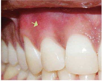
Figure 1 Labial view with draining sinus tract (yellow arrow) associated with maxillary right lateral incisor.
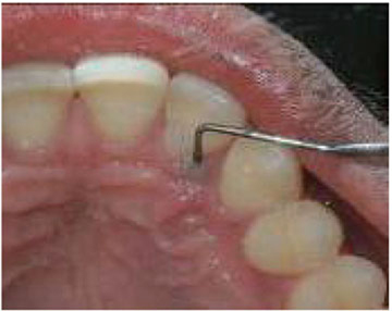
Figure 2 Palatal view (probing depth, 15 mm).

Figure 3 Preoperative intraoral periapical radiograph.
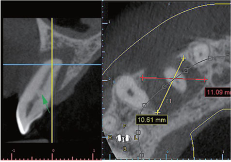
Figure 4 Preoperative cone beam computed tomography images. Green arrow indicates nonembeded pulp stone.
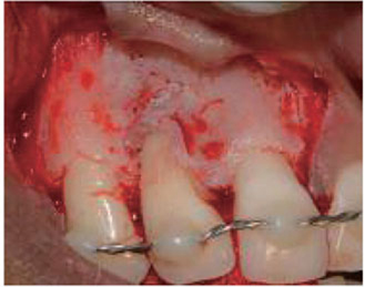
Figure 5 Reflection of mucoperiosteal flap.

Figure 6 After complete removal of granulation tissue. Yellow arrow indicates extrusion of approximately 1 mm of gutta-percha from the apex.
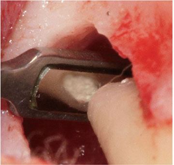
Figure 7 Mineral trioxide aggregate restoration after retropreparation.
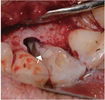
Figure 8 Glass ionomer cement restoration in the palatoradicular groove. White arrow indicates glass ionomer restoration.
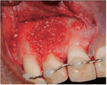
Figure 9 Bone defect filled with mixture of demineralized freeze-dried bone allograft and platelet rich fibrin.
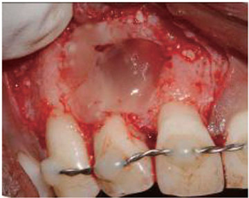
Figure 10 Platelet rich fibrin used as membrane.
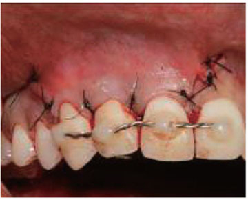
Figure 11 Suture to achieve primary closure.
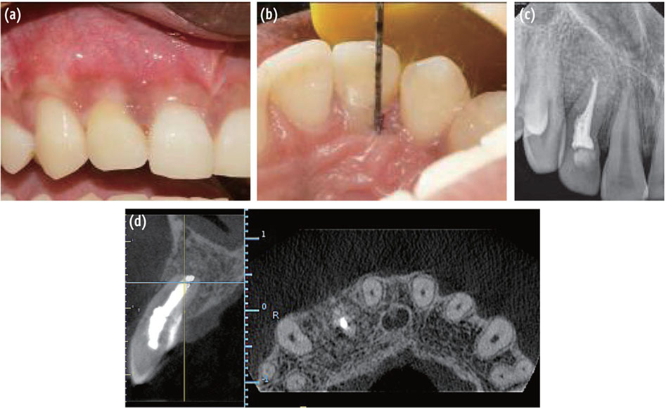
Figure 12 Clinical photographs (a, b) and radiographs (c, d) at 12 month follow up. (a) Labial view; (b) Palatal view; (c) Intraoral periapical radiograph; (d) Cone beam computed tomography.

Figure 13 Photomicrograph of the lesion showing surface squamous epithelium with arcading type of proliferation (blue arrow) and chronic inflammatory cell infiltration in connective tissue (Haematoxylin and eosin stain, ×10).
Reference
-
1. Darby I, Curtis M. Microbiology of periodontal disease in children and young adults. Periodontol 2000. 2001; 26:33–53.
Article2. Kovács V, Tihanyi D, Gera I. The incidence of local plaque retentive factors in chronic periodontitis. Fogorv Sz. 2007; 100:295–300.3. Simon JH, Glick DH, Frank AL. Predictable endodontic and periodontic failures as a result of radicular anomalies. Oral Surg Oral Med Oral Pathol. 1971; 31:823–826.
Article4. Ennes JP, Lara VS. Comparative morphological analysis of the root developmental groove with the palato-gingival groove. Oral Dis. 2004; 10:378–382.
Article5. Peikoff MD, Perry JB, Chapnick LA. Endodontic failure attributable to a complex radicular lingual groove. J Endod. 1985; 11:573–577.
Article6. Kogon SL. The prevalence, location and conformation of palato-radicular grooves in maxillary incisors. J Periodontol. 1986; 57:231–234.
Article7. Assaf ME, Roller N. The cingulo-radicular groove: its significance and management-two cases reports. Compendium. 1992; 13:94. 96. 98 passim.8. Lara VS, Consolaro A, Bruce RS. Macroscopic and Microscopic analysis of the palato-gingival groove. J Endod. 2000; 26:345–350.
Article9. Dietrich T, Zunker P, Dietrich D, Bernimoulin JP. Apicomarginal defects in periradicular surgery: classification and diagnostic aspects. Oral Surg Oral Med Oral Pathol Oral Radiol Endod. 2002; 94:233–239.
Article10. Goyal B, Tewari S, Duhan J, Sehgal PK. Comparative evaluation of platelet-rich plasma and guided tissue regeneration membrane in the healing of apicomarginal defects: a clinical study. J Endod. 2011; 37:773–780.
Article11. Abbott PV, Salgado JC. Strategies for the endodontic management of concurrent endodontic and periodontal diseases. Aust Dent J. 2009; 54:S70–S85.
Article12. Estrela C, Bueno MR, Azevedo BC, Azevedo JR, Pécora JD. A new periapical index based on cone beam computed tomography. J Endod. 2008; 34:1325–1331.
Article13. Oh SL, Fouad AF, Park SH. Treatment strategy for guided tissue regeneration in combined endodontic-periodontal lesions: case report and review. J Endod. 2009; 35:1331–1336.
Article14. Rud J, Andreasen JO, Jensen JE. Radiographic criteria for the assessment of healing after endodontic surgery. Int J Oral Surg. 1972; 1:195–214.
Article15. Schulz A, Hilgers RD, Niedermeier W. The effect of splinting of teeth in combination with reconstructive periodontal surgery in humans. Clin Oral Investig. 2000; 4:98–105.
Article16. Sekhar LC, Koganti VP, Shankar BR, Gopinath A. A comparative study of temporary splints: bonded polyethylene fiber reinforcement ribbon and stainless steel wire + composite resin splint in the treatment of chronic periodontitis. J Contemp Dent Pract. 2011; 12:343–349.
Article17. Ballal NV, Jothi V, Bhat KS, Bhat KM. Salvaging a tooth with a deep palatogingival groove: an endo-perio treatment - a case report. Int Endod J. 2007; 40:808–817.
Article18. Kishan KV, Hegde V, Ponnappa KC, Girish TN, Ponappa MC. Management of palato-radicular groove in a maxillary lateral incisor. J Nat Sci Biol Med. 2014; 5:178–181.19. Skoglund A, Persson G. A follow-up study of apicoectomized teeth with total loss of the buccal bone plate. Oral Surg Oral Med Oral Pathol. 1985; 59:78–81.
Article20. Abramowitz PN, Rankow H, Trope M. Multidisciplinary approach to apical surgery in conjunction with the loss of buccal cortical plate. Oral Surg Oral Med Oral Pathol. 1994; 77:502–506.
Article21. Gutmann JL, Harrison JW. Surgical Endodontics. 1st ed. Chennai: All India Publishers and Distributors;1999. p. 338.22. Taschieri S, Rosano G, Weinstein T, Bortolin M, Del Fabbro M. Treatment of through-and-through bone lesion using autologous growth factors and xenogeneic bone graft: a case report. Oral Maxillofac Surg. 2012; 16:57–64.
Article23. Attam K, Tiwary R, Talwar S, Lamba AK. Palatogingival groove: endodontic-periodontal management-case report. J Endod. 2010; 36:1717–1720.
Article24. Shivashankar VY, Johns DA, Vidyanath S, Sam G. Combination of platelet rich fibrin, hydroxyapatite and PRF membrane in the management of large inflammatory periapical lesion. J Conserv Dent. 2013; 16:261–264.
Article25. Choukroun J, Diss A, Simonpieri A, Girard MO, Schoeffler C, Dohan SL, Anthony J, Dohan J, Mouhyi J, Dohan DM. Platelet-rich fibrin (PRF): a second-generation platelet concentrate. Part IV. clinical effects on tissue healing. Oral Surg Oral Med Oral Pathol Oral Radiol Endod. 2006; 101:e56–e60.
Article26. Pecora G, Baek SH, Rethnam S, Kim S. Barrier membrane techniques in endodontic microsurgery. Dent Clin North Am. 1997; 41:585–602.27. Gomes SC, Miranda LA, Soares I, Oppermann RV. Clinical and Histologic Evaluation of the Periodontal Response to Restorative Procedures in the Dog. Int J Periodontics Restorative Dent. 2005; 25:39–47.28. Forero-López J, Gamboa-Martínez L, Pico-Porras L, Niño-Barrera JL. Surgical management with intentional replantation on a tooth with palato-radicular groove. Restor Dent Endod. 2015; 40:166–171.
Article29. Naik M, de Ataide Ide N, Fernandes M, Lambor R. Treatment of combined endodontic: periodontic lesion by sealing of palato-radicular groove using biodentine. J Conserv Dent. 2014; 17:594–597.
Article30. Zucchelli G, Mele M, Checchi L. The papilla amplification flap for the treatment of a localized periodontal defect associated with a palatal groove. J Periodontol. 2006; 77:1788–1796.
Article31. Rachana D, Nadig P, Nadig G. The palatal groove: application of computed tomography in its detection - a case report. J Conserv Dent. 2007; 10:83–88.
Article32. Rajput A, Talwar S, Chaudhary S, Khetarpal A. Successful management of pulpo-periodontal lesion in maxillary lateral incisor with palatogingival groove using CBCT scan. Indian J Dent Res. 2012; 23:415–418.
Article
- Full Text Links
-
- Actions
-
Cited
- CITED
-
- Close
- Share
-
- Similar articles
-
- Non-destructive management of white spot lesions by using tooth jewelry
- Clinical crown lengthening procedure using surgical extrusion in esthetic region
- Outcome Evaluation of an Immediately Placed Maxillary Anterior Single-Tooth Implant Using Objective Esthetic Criteria: Case Report
- Esthetic enhancement of a traumatized anterior tooth with a combination of forced eruption and tooth alignment: a case report
- Combined application of roll flap and combination onlay-interpositional graft to enhance esthetics of maxillary anterior fixed partial denture: A case report
