Combined application of roll flap and combination onlay-interpositional graft to enhance esthetics of maxillary anterior fixed partial denture: A case report
- Affiliations
-
- 1Department of Prosthodontics, College of Dentistry, Wonkwang University, Daejeon, Republic of Korea. scoh@wku.ac.kr
- KMID: 2393198
- DOI: http://doi.org/10.4047/jap.2016.8.1.70
Abstract
- In the maxillary anterior region, reconstruction of the localized alveolar ridge defect is very important in enhancing the esthetics of fixed partial denture. A 40-year-old female patient presented with a chief complaint of the inconvenience and unesthetic problem of 3-unit maxillary anterior prosthesis due to alveolar ridge resorption. After removal of old prosthesis, intraoral examination revealed moderate (buccolingually 4 mm) ridge deficiency in missing tooth region, leading to the diagnosis of Class I alveolar ridge defect. One of the reconstruction techniques to overcome this problem might be a technique that combines two types of soft tissue augmentation techniques. The purpose of this paper was to demonstrate the new combined technique of roll flap and combination onlay-interpositional graft utilized to acquire sufficient dimension of recipient area by one time of operation and to present the esthetic improvement of fixed partial denture by using this procedure in case of maxillary anterior localized ridge defect.
Keyword
MeSH Terms
Figure
-
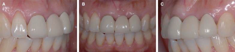
Fig. 1 Pretreatment views of 3-unit fixed partial denture at the time of initial examination. (A, C) Lateral views and (B) frontal view showed the labial concavity of gingiva and the space between pontic and underlying gingiva.
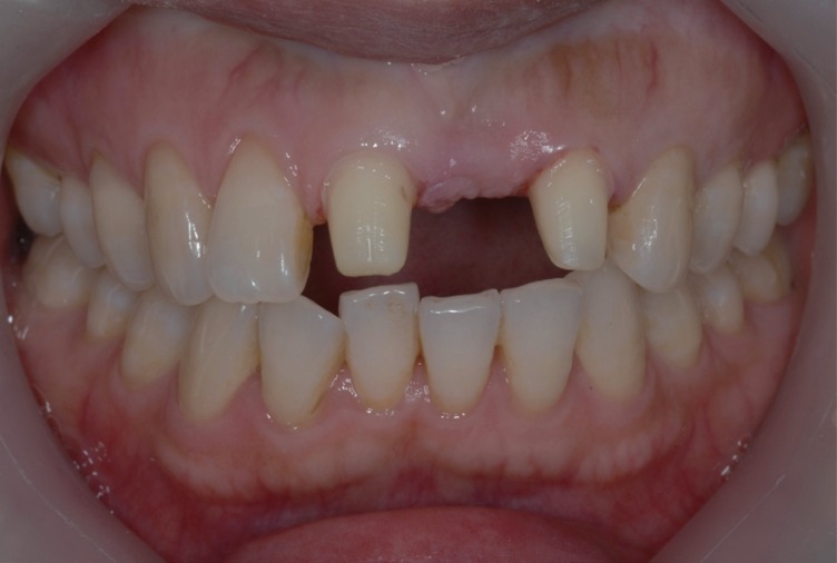
Fig. 2 Labial view of a Class I ridge defect. Note moderate labial ridge deficiency with normal ridge height of missing tooth region.
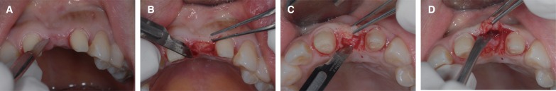
Fig. 3 Roll flap procedure (A) a partial-thickness crestal incision was made mesiodistally at the height of ridge. (B) the gingival surface was de-epithelialized in a trapezoid on palatal side of the ridge. (C) the full-thickness rectangular pedicle with de-epithelialized connective tissue was made by a No. 15 scalpel blade. (D) The de-epithelialized connective tissue pedicle raised from palate using sharp dissection was rolled and placed in the pocket on the labial surface of the deformed ridge.
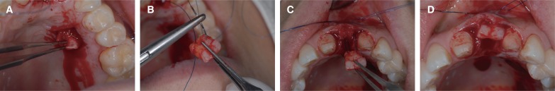
Fig. 4 Combination onlay-interpositional graft procedure. (A) The donor site was prepared on the premolar palatal area. (B) The trapezoidal shaped transplant was made of the de-epithelialized and epithelialized part. (C) The transplant was immediately fixed with two separate sutures prepared in advance on both ends of the bottom of the pouch. (D) The epithelialized section of the graft was positioned above the surface of the surrounding tissue at the crest of the ridge.
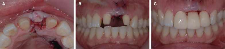
Fig. 5 Sutures and immediate placement of provisional restoration. (A) Occlusal view and (B) labial view showed both apical single-knot sutures to stabilize the transplant. (C) Provisional prosthesis with ovate pontic was directly modified and placed in position.
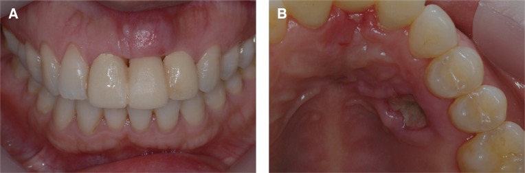
Fig. 6 Healing state after 1 week post-surgery. (A) Labial view of recipitent site and (B) occlusal view of donor site showed good healing at 1 week post-surgery.
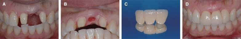
Fig. 7 Recipient site and delivery of final restoration at 7 weeks post-surgery. (A) Labial view and (B) occlusal view showed good ovate seat but the trace of crestal incision line was remained at mesial side of lateral incisor. (C) Final prosthesis was zirconial all-ceramic restoration with ovate pontic. (D) Final restoration showed good emergence profile and no interference in function.
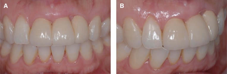
Fig. 8 Labial view of final prosthesis after 18 months post-surgery. (A) Frontal view and (B) lateral view showed natural-looking emergence profile, stable alveolar ridge, and harmonious color.
Reference
-
1. Lazzara RJ. Managing the soft tissue margin: the key to implant aesthetics. Pract Periodontics Aesthet Dent. 1993; 5:81–88. PMID: 8219171.2. Seibert JS, Salama H. Alveolar ridge preservation and reconstruction. Periodontol 2000. 1996; 11:69–84. PMID: 9567959.
Article3. Seibert JS. Ridge augmentation to enhance esthetics in fixed prosthetic treatment. Compendium. 1991; 12:548. 550. 552 passim. PMID: 1809510.4. Yildirim M, Hanisch O, Spiekermann H. Simultaneous hard and soft tissue augmentation for implant-supported single-tooth restorations. Pract Periodontics Aesthet Dent. 1997; 9:1023–1031. quiz 1032PMID: 9573855.5. Studer S, Naef R, Schärer P. Adjustment of localized alveolar ridge defects by soft tissue transplantation to improve mucogingival esthetics: a proposal for clinical classification and an evaluation of procedures. Quintessence Int. 1997; 28:785–805. PMID: 9477870.6. Abrams L. Augmentation of the deformed residual edentulous ridge for fixed prosthesis. Compend Contin Educ Gen Dent. 1980; 1:205–213. PMID: 6950834.7. Scharf DR, Tarnow DP. Modified roll technique for localized alveolar ridge augmentation. Int J Periodontics Restorative Dent. 1992; 12:415–425. PMID: 1343013.8. Seibert JS. Reconstruction of deformed, partially edentulous ridges, using full thickness onlay grafts. Part I. Technique and wound healing. Compend Contin Educ Dent. 1983; 4:437–453. PMID: 6578906.9. Coslet JG, Rosenberg ES, Tisot R. The free autogenous gingival graft. Dent Clin North Am. 1980; 24:651–682. PMID: 7000559.10. Orth CF. A modification of the connective tissue graft procedure for the treatment of type II and type III ridge deformities. Int J Periodontics Restorative Dent. 1996; 16:266–277. PMID: 9084312.11. Langer B, Calagna L. The subepithelial connective tissue graft. J Prosthet Dent. 1980; 44:363–367. PMID: 6931898.
Article12. Garber D, Rosenberg E. The edentulous ridge in fixed prosthodontics. Compend Contin Educ Dent. 1981; 2:212–223. PMID: 6950860.13. Seibert JS, Louis JV. Soft tissue ridge augmentation utilizing a combination onlay-interpositional graft procedure: a case report. Int J Periodontics Restorative Dent. 1996; 16:310–321. PMID: 9242099.14. Stimmelmayr M, Allen EP, Reichert TE, Iglhaut G. Use of a combination epithelized-subepithelial connective tissue graft for closure and soft tissue augmentation of an extraction site following ridge preservation or implant placement: description of a technique. Int J Periodontics Restorative Dent. 2010; 30:375–381. PMID: 20664839.15. Allen EP, Gainza CS, Farthing GG, Newbold DA. Improved technique for localized ridge augmentation. A report of 21 cases. J Periodontol. 1985; 56:195–199. PMID: 2987473.
Article16. Perenack J, Wood RJ, Block MS, Gardiner D. Determination of subepithelial connective tissue graft thickness in the dog. J Oral Maxillofac Surg. 2002; 60:415–421. PMID: 11928100.
Article17. Oliver RC, Löe H, Karring T. Microscopic evaluation of the healing and revascularization of free gingival grafts. J Periodontal Res. 1968; 3:84–95. PMID: 4249992.
Article18. Wiskott HWA. Fixed Prosthodontics: Principles and clinics. Great Britian: Quintessence;2011. p. 243.
- Full Text Links
-
- Actions
-
Cited
- CITED
-
- Close
- Share
-
- Similar articles
-
- Implant-assisted removable partial denture in a maxillary edentulous patient: A case report
- Multidisciplinary approach of the problem of unaesthetic implants in the maxillary anterior dentition
- Resin-bonded fixed partial denture using In-Ceram and Targis-Ventris system
- Distal-extension removable partial denture with anterior implant supported fixed prostheses in a maxillary edentulous patient: Case report
- An implant-supported removable partial denture for a patient with post-inflammatory scar contracture caused by burn complications: a clinical report
