Restor Dent Endod.
2014 May;39(2):132-136.
Endodontic management of a mandibular second molar with radix entomolaris: a case report
- Affiliations
-
- 1Department of Conservative Dentistry and Endodontics, Faculty of Dental Sciences, Sri Ramachandra University, Porur, Chennai, India. hannahrosaline@gmail.com
- 2Department of Conservative Dentistry and Endodontics, JKK Nataraja Dental College, Komarapalayam, Erode, India.
Abstract
- The presence of radix entomolaris (RE) in a mandibular first molar is a common occurrence in certain ethnic groups, but the presence of RE in a mandibular second molar is a rare occurrence. In the present case, RE was identified from preoperative radiographs and confirmed using cone-beam computed tomography (CBCT). The access cavity was modified to locate the RE. Cleaning and shaping were performed with nickel-titanium rotary instruments. Obturation was completed with gutta-percha cones using AH Plus (Dentsply Detrey GmbH) as sealer. From the CBCT axial images, the RE was determined to have a Type III curvature by the De Moor classification, Type B separate RE by the Carlsen and Alexandersen classification, and radiographically, a Type i image by the Wang classification. The presence of RE in the mandibular second molar makes it essential to anticipate and treat the distolingual root canal. This case report highlights the usefulness of CBCT for assessing RE in the mandibular second molar, which can help the clinician in making a confirmatory diagnosis and assessing the morphology of the root canal.
MeSH Terms
Figure
-
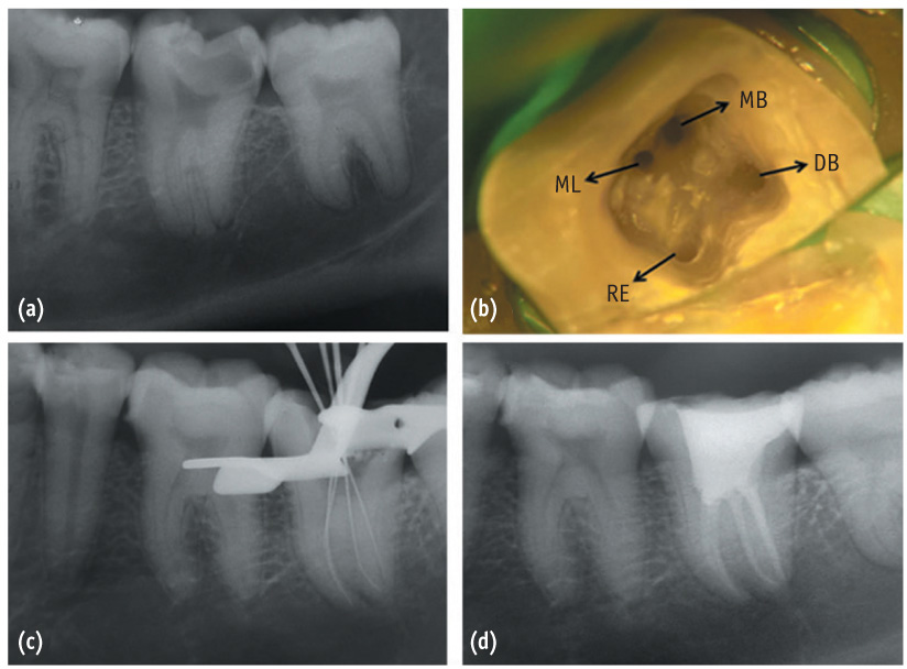
Figure 1 (a) Preoperative radiograph of #37; (b) Access opening showing the presence of RE, MB, ML, and DB canal orifices; (c) Working length radiograph; (d) Postoperative radiograph. RE, radix entomolaris; MB, mesio buccal; ML, mesio lingual; DB, disto buccal.

Figure 2 CBCT images of #37 confirming the presence of a separate RE (arrow) (a) At the cervical level; (b) At the apical level. (c) The CBCT enlarged section at cervical level. (d) CBCT sagittal section showing type III curvature.
Reference
-
1. Schumacher C. Endodontic treatment of a mandibular first molar with radix entomolaris: a case report. ENDO (Lond Engl). 2008; 2:301–304.2. Carabelli G. Systematisches Handbuch Der Zahnheikunde. 2nd ed. Vienna: Braumuller and Seidel;1844. p. 114.3. Calberson FL, De Moor RJ, Deroose CA. The radix entomolaris and paramolaris: clinical approach in endodontics. J Endod. 2007; 33:58–63.
Article4. Manning SA. Root canal anatomy of mandibular second molars. Part I. Int Endod J. 1990; 23:34–39.
Article5. Neelakantan P, Subbarao C, Subbarao CV, Ravindranath M. Root and canal morphology of mandibular second molars in an Indian population. J Endod. 2010; 36:1319–1322.
Article6. Garg AK, Tewari RK, Kumar A, Hashmi SH, Agrawal N, Mishra SK. Prevalence of three-rooted mandibular permanent first molars among the Indian population. J Endod. 2010; 36:1302–1306.
Article7. Turner CG 2nd. Three-rooted mandibular first permanent molars and the question of American Indian origins. Am J Phys Anthropol. 1971; 34:229–241.
Article8. Loh HS. Incidence and features of three-rooted permanent mandibular molars. Aust Dent J. 1990; 35:434–437.
Article9. de Souza-Freitas JA, Lopes ES, Casati-Alvares L. Anatomic variations of lower first permanent molar roots in two ethnic groups. Oral Surg Oral Med Oral Pathol. 1971; 31:274–278.
Article10. Skidmore AE, Bjorndal AM. Root canal morphology of the human mandibular first molar. Oral Surg Oral Med Oral Pathol. 1971; 32:778–784.
Article11. Steelman R. Incidence of an accessory distal root on mandibular first permanent molars in Hispanic children. ASDC J Dent Child. 1986; 53:122–123.12. Sperber GH, Moreau JL. Study of the number of roots and canals in Senegalese first permanent mandibular molars. Int Endod J. 1998; 31:117–122.
Article13. Tratman EK. Three-rooted lower molars in man and their racial distribution. Br Dent J. 1938; 64:264–274.14. Tachibana H, Matsumoto K. Applicability of X-Ray computerized tomography in endodontics. Endod Dent Traumatol. 1990; 6:16–20.
Article15. Nair MK, Nair UP. Digital and advanced imaging in endodontics: a review. J Endod. 2007; 33:1–6.
Article16. Matherne RP, Angelopoulos C, Kulild JC, Tira D. Use of cone-beam computed tomography to identify root canal systems in vitro. J Endod. 2008; 34:87–89.
Article17. Song JS, Choi HJ, Jung IY, Jung HS, Kim SO. The prevalence and morphologic classification of distolingual roots in the mandibular molars in a Korean population. J Endod. 2010; 36:653–657.
Article18. Schäfer E, Breuer D, Janzen S. The prevalence of three-rooted mandibular permanent first molars in a German population. J Endod. 2009; 35:202–205.
Article19. Reichart PA, Metah D. Three-rooted permanent mandibular first molars in the Thai. Community Dent Oral Epidemiol. 1981; 9:191–192.
Article20. Gu Y, Lu Q, Wang H, Ding Y, Wang P, Ni L. Root canal morphology of permanent three-rooted mandibular first molars-part I: pulp floor and root canal system. J Endod. 2010; 36:990–994.
Article21. De Moor RJ, Deroose CA, Calberson FL. The radix entomolaris in mandibular first molars: an endodontic challenge. Int Endod J. 2004; 37:789–799.
Article22. Wang Q, Yu G, Zhou XD, Peters OA, Zheng QH, Huang DM. Evaluation of X-ray projection angulation for successful radix entomolaris diagnosis in mandibular first molars in vitro. J Endod. 2011; 37:1063–1068.
Article23. Carlsen O, Alexandersen V. Radix entomolaris: identification and morphology. Scand J Dent Res. 1990; 98:363–373.
Article24. Patel S, Dawood A, Whaites E, Pitt Ford T. New dimensions in endodontic imaging: part 1. Conventional and alternative radiographic systems. Int Endod J. 2009; 42:447–462.
Article25. Karanxha L, Kim HJ, Hong SO, Lee W, Kim PS, Min KS. Endodontic management of a C-shaped maxillary first molar with three independent buccal root canals by using cone-beam computed tomography. Restor Dent Endod. 2012; 37:175–179.
Article26. Abella F, Patel S, Durán-Sindreu F, Mercadé M, Roig M. Mandibular first molars with disto-lingual roots: review and clinical management. Int Endod J. 2012; 45:963–978.
Article27. Lee MH, Ha JH, Jin MU, Kim YK, Kim SK. Endodontic treatment of maxillary lateral incisors with anatomical variations. Restor Dent Endod. 2013; 38:253–257.
Article
- Full Text Links
-
- Actions
-
Cited
- CITED
-
- Close
- Share
-
- Similar articles
-
- Asymmetry in mesial root number and morphology in mandibular second molars: a case report
- Radix mesiolingualis and radix distolingualis: a case report of a tooth with an unusual morphology
- The prevalence of radix molaris in the mandibular first molars of a Saudi subpopulation based on cone-beam computed tomography
- Cone beam computed tomography findings of ectopic mandibular third molar in the mandibular condyle: report of a case
- Management of failed periodontal surgical intervention for a furcal lesion with a nonsurgical endodontic approach
