Restor Dent Endod.
2014 May;39(2):120-125.
Biodentine-a novel dentinal substitute for single visit apexification
- Affiliations
-
- 1Department of Conservative Dentistry and Endodontics, Kanti Devi Dental College and Hospital, Mathura, Uttar Pradesh, India. gurudutt_nayak@ hotmail.com
Abstract
- Use of an apical plug in management of cases with open apices has gained popularity in recent years. Biodentine, a new calcium silicate-based material has recently been introduced as a dentine substitute, whenever original dentine is damaged. This case report describes single visit apexification in a maxillary central incisor with necrotic pulp and open apex using Biodentine as an apical barrier, and a synthetic collagen material as an internal matrix. Following canal cleaning and shaping, calcium hydroxide was placed as an intracanal medicament for 1 mon. This was followed by placement of small piece of absorbable collagen membrane beyond the root apex to serve as matrix. An apical plug of Biodentine of 5 mm thickness was placed against the matrix using pre-fitted hand pluggers. The remainder of canal was back-filled with thermoplasticized gutta-percha and access cavity was restored with composite resin followed by all-ceramic crown. One year follow-up revealed restored aesthetics and function, absence of clinical signs and symptoms, resolution of periapical rarefaction, and a thin layer of calcific tissue formed apical to the Biodentine barrier. The positive clinical outcome in this case is encouraging for the use of Biodentine as an apical plug in single visit apexification procedures.
Keyword
MeSH Terms
Figure
-

Figure 1 (a) Preoperative photograph showing Elli's Class IV fracture and discolouration in relation to 21; (b) Postoperative photograph showing tooth restored with an all-ceramic crown.
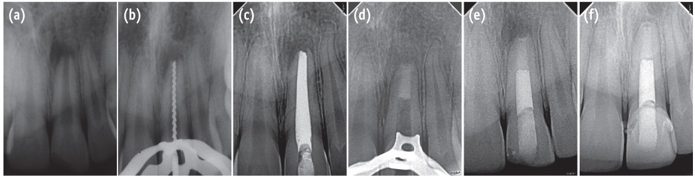
Figure 2 (a) Preoperative radiograph showing tooth 21 with open apices and periapical radiolucency; (b) Working length radiograph; (c) Calcium hydroxide as intracanal medicament; (d) Radiograph confirming the placement of Biodentine apical plug compacted against the barrier material; (e) Backfill performed using thermoplasticized gutta-percha; (f) One year follow-up radiograph showing resolution of periapical rarefaction and a thin layer of calcific tissue formed apical to the Biodentine barrier.
Reference
-
1. Bhasker SN. Orban's oral histology and embryology. 11th ed. St. Louis: Mosby-Year Book;1991. p. 382.2. American Association of Endodontists: Glossary of endodontic terms. updated 2013 Nov 6. Available from: http://www.aae.org/glossary.3. Sheehy EC, Roberts GJ. Use of calcium hydroxide for apical barrier formation and healing in non-vital immature permanent teeth: a review. Br Dent J. 1997; 183:241–246.
Article4. Felippe MC, Felippe WT, Marques MM, Antoniazzi JH. The effect of renewal of calcium hydroxide paste on the apexification and periapical healing of teeth with incomplete root formation. Int Endod J. 2005; 38:436–442.
Article5. Metzger Z, Solomonov M, Mass E. Calcium hydroxide retention in wide root canals with flaring apices. Dent Traumatol. 2001; 17:86–92.
Article6. Andreasen JO, Farik B, Munksgaard EC. Long-term calcium hydroxide as a root canal dressing may increase risk of root fracture. Dent Traumatol. 2002; 18:134–137.
Article7. Binnie WH, Rowe AH. A histological study of periapical tissues of incompletely formed pulpless teeth filled with calcium hydroxide. J Dent Res. 1973; 52:1110–1116.
Article8. Roberts SC Jr, Brilliant JD. Tricalcium phosphate as an adjunct to apical closure in pulpless permanent teeth. J Endod. 1975; 1:263–269.
Article9. Rossmeisl R, Reader A, Melfi R, Marquard J. A study of freeze-dried (lyophilized) cortical bone used as an apical barrier in adult monkey teeth. J Endod. 1982; 8:219–226.
Article10. Rossmeisl R, Reader A, Melfi R, Marquard J. A study of freeze-dried (lyophilized) dentin used as an apical barrier in adult monkey teeth. Oral Surg Oral Med Oral Pathol. 1982; 53:303–310.
Article11. Nevins A, Finkelstein F, Laporta R, Borden BG. Induction of hard tissue into pulpless open-apex teeth using collagen-calcium phosphate gel. J Endod. 1978; 4:76–81.
Article12. Eleazer PD, McDonald TW, Sinai IH, Fantasia JE, Michelich RJ, Yagiela JA. Proplast as an apical barrier in root canal therapy. J Endod. 1984; 10:487–490.
Article13. Torabinejad M, Chivian N. Clinical applications of mineral trioxide aggregate. J Endod. 1999; 25:197–205.
Article14. Lemon RR. Nonsurgical repair of perforation defects. Internal matrix concept. Dent Clin North Am. 1992; 36:439–457.15. Bargholz C. Perforation repair with mineral trioxide aggregate: a modified matrix concept. Int Endod J. 2005; 38:59–69.
Article16. Mesimeris V, Sade E, Baer PN. Calcium sulfate as a biodegradable barrier membrane: a preliminary report on the "Surgiplast" technique. Periodontal Clin Investig. 1995; 17:13–16.17. Rudagi KB, Rudagi B. One-step apexification in immature tooth using grey mineral trioxide aggregate as an apical barrier and autologus platelet rich fibrin membrane as an internal matrix. J Conserv Dent. 2012; 15:196–199.
Article18. Parirokh M, Torabinejad M. Mineral trioxide aggregate: a comprehensive literature review - Part I: chemical, physical, and antibacterial properties. J Endod. 2010; 36:16–27.
Article19. Torabinejad M, Parirokh M. Mineral trioxide aggregate: a comprehensive literature review - Part II: leakage and biocompatibility investigations. J Endod. 2010; 36:190–202.
Article20. Parirokh M, Torabinejad M. Mineral trioxide aggregate: a comprehensive literature review - Part III: clinical applications, drawbacks, and mechanism of action. J Endod. 2010; 36:400–413.
Article21. Chang SW. Chemical characteristics of mineral trioxide aggregate and its hydration reaction. Restor Dent Endod. 2012; 37:188–193.
Article22. Septodont. Biodentine - Active Biosilicate Technology, scientific file. Saint-Maur-des-Fossés Cedex, France: R&D Department, Septodont;2010.23. Villat C, Grosgogeat B, Seux D, Farge P. Conservative approach of a symptomatic carious immature permanent tooth using a tricalcium silicate cement (Biodentine): a case report. Restor Dent Endod. 2013; 38:258–262.
Article24. Garrault S, Behr T, Nonat A. Formation of the C-S-H Layer during early hydration of tricalcium silicate grains with different sizes. J phys chem B. 2006; 110:270–275.
Article25. Pradelle-Plasse N, Tran XV, Colon P. VI-2-1. Physico chemical properties. In : Goldberg M, editor. Biocompatibility or cytotoxic effects of dental composites. Oxford: Coxmoor Publishing Co.;2009. p. 184–194.26. Bachoo IK, Seymour D, Brunton P. A biocompatible and bioactive replacement for dentine: is this a reality? The properties and uses of a novel calcium-based cement. Br Dent J. 2013; 214:E5.
Article27. Bachoo IK, Seymour D, Brunton P. Clinical case reports using a novel calcium-based cement. Br Dent J. 2013; 214:61–64.
Article28. Camilleri J. Characterization of hydration products of mineral trioxide aggregate. Int Endod J. 2008; 41:408–417.
Article29. Tay FR, Pashley DH, Rueggeberg FA, Loushine RJ, Weller RN. Calcium phosphate phase transformation produced by the interaction of the portland cement component of white mineral trioxide aggregate with a phosphatecontaining fluid. J Endod. 2007; 33:1347–1351.
Article30. Goldberg M. Chapter VI. Emerging trends in (bio)material researches. Biocompatibility or cytotoxic effects of dental composites. Oxford: Coxmoor Publishing Co.;2009. p. 181–203.31. Han L, Okiji T. Uptake of calcium and silicon released from calcium silicate-based endodontic materials into root canal dentine. Int Endod J. 2011; 44:1081–1087.
Article32. Benenati FW, Roane JB, Biggs JT, Simon JH. Recall evaluation of iatrogenic root perforations repaired with amalgam and gutta-percha. J Endod. 1986; 12:161–166.
Article33. Thibodeau B, Trope M. Pulp revascularization of a necrotic infected immature permanent tooth: case report and review of the literature. Pediatr Dent. 2007; 29:47–50.
- Full Text Links
-
- Actions
-
Cited
- CITED
-
- Close
- Share
-
- Similar articles
-
- Outcome of Regenerative Endodontic Treatment for an Avulsed Immature Permanent Tooth: A Case Report
- One-visit Apexification Using MTA and Reattachment of a Crown-root Fractured Tooth with Severe Coronal Damage: A Case Report
- A review of the regenerative endodontic treatment procedure
- Observation of an extracted premolar 2.5 years after mineral trioxide aggregate apexification using micro-computed tomography
- Interface between calcium silicate cement and adhesive systems according to adhesive families and cement maturation
