Vital tooth with periapical lesion: spontaneous healing after conservative treatment
- Affiliations
-
- 1Department of Conservative Dentistry, Yonsei University College of Dentistry, Seoul, Korea. sunghopark@yuhs.ac
Abstract
- It is often presumed that apical periodontitis follows total pulp necrosis, and consequently root canal treatment is commonly performed. Periapical lesion development is usually caused by bacteria and its byproduct which irritate pulp, develop pulpitis, and result in necrosis through an irreversible process. Afterwards, apical periodontitis occurs. This phenomenon is observed as an apical radiolucency in radiographic view. However, this unusual case presents a spontaneous healing of periapical lesion, which has developed without pulp necrosis in a vital tooth, through conservative treatment.
MeSH Terms
Figure
-

Figure 1 Preoperative periapical view and clinical photograph. (a) Periapical radiolucency on #35; (b) Cervical abrasion.

Figure 2 Preoperative panorama view. Periapical radiolucency was observed at the apex of mandibular left second premolar.

Figure 3 Periapical view taken 2 years ago.

Figure 4 Location of mental foramens (dotted line) and periapical radiolucency (arrow).

Figure 5 Periapical view and clinical photograph after class V Resin filling.

Figure 6 Three-month follow up. (a) Periapical view. Distal proximal caries was detected; (b) Photograph taken after class II resin filling.

Figure 7 Six-month follow up. Periapical view.

Figure 8 Nine-month follow up. Periapical view.
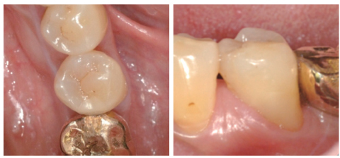
Figure 9 Fourteen-month follow up. Clinical photographs.

Figure 10 Fourteen-month follow up. Horizontal shift periapical view.

Figure 11 One year before implant surgery: Note that the posterior area of mandibular left segment was edentulous.
Reference
-
1. Kim IS, Kim SG, Kim YK, Kim JD. Position of the mental foramen in a Korean population: a clinical and radiographic study. Implant Dent. 2006. 15:404–411.
Article2. Resnick CM, Novelline RA. Cemento-osseous dysplasia, a radiological mimic of periapical dental abscess. Emerg Radiol. 2008. 15:367–374.
Article3. Romanos GE, Froum S, Costa-Martins S, Meitner S, Tarnow DP. Implant periapical lesions: etiology and treatment options. J Oral Implantol. 2011. 37:53–63.
Article4. Kovacević M, Tamarut T, Jonjić N, Braut A. The transition from pulpitis to periapical periodontitis in dogs' teeth. Aust Endod J. 2008. 34:12–18.
Article5. Khayat BG, Byers MR, Taylor PE, Mecifi K, Kimberly CL. Responses of nerve fibers to pulpal inflammation and periapical lesions in rat molars demonstrated by calcitonin gene-related peptide immunocytochemistry. J Endod. 1988. 14:577–587.
Article6. Byers MR, Taylor PE, Khayat BG, Kimberly CL. Effects of injury and inflammation on pulpal and periapical nerves. J Endod. 1990. 16:78–84.
Article7. Caviedes-Bucheli J, Muñoz HR, Azuero-Holguín MM, Ulate E. Neuropeptides in dental pulp: the silent protagonists. J Endod. 2008. 34:773–788.
Article8. Stashenko P, Teles R, D'Souza R. Periapical inflammatory responses and their modulation. Crit Rev Oral Biol Med. 1998. 9:498–521.
Article
- Full Text Links
-
- Actions
-
Cited
- CITED
-
- Close
- Share
-
- Similar articles
-
- Diagnosis of periapical cemental dysplasia
- Endodontic management of central incisor associated with large periapical lesion and fused supernumerary root: a conservative approach
- A retrospective study of the intentionally replanted mandibular second molars with C-shaped root canal configurations
- Partial pulp necrosis caused by excessive orthodontic force
- The significance of diagnosis and treatment planning in periapical lesion overfilled with calcium hydroxide paste
