Feasibility Study of the microDiamond Detector for Measurement of Small Field Photon Beam
- Affiliations
-
- 1Department of Radiation Oncology, College of Medicine, Inha University, Incheon, Korea. hyundohuh@gmail.com
- 2Research Institute of Radiological and Medical Sciences, Korea Institute of Radiological and Medical Sciences, Seoul, Korea.
- 3Department of Radiation Oncology, College of Medicine, Kwandong University, Gangneung, Korea.
- 4Department of Radiation Oncology, College of Medicine, Soonchunhyang University, Cheonan, Korea.
- 5Department of Radiation Oncology, College of Medicine, Kyunghee University, Seoul, Korea.
- 6Department of Radiation Oncology, College of Medicine, Hanyang University, Seoul, Korea.
- KMID: 2315765
- DOI: http://doi.org/10.14316/pmp.2014.25.4.255
Abstract
- The dosimetry of very small fields is challenging for several reasons including a lack of lateral electronic equilibrium, large dose gradients, and the size of detector in respect to the field size. The objective of this work was to evaluate the suitability of a new commercial synthetic diamond detector, namely, the PTW 60019 microDiamond, for the small field dosimetry in cyberknife photon beams of 6 different collimator size (from 5 mm to 30 mm). Measurements included dose linearity, dose rate dependence, output factors (OF), percentage depth doses (PDD) and off center ratio (OCR). The results were compared to those of pinpoint ionization chamber, diamond detector, microLion liquid Ionization chamber and diode detector. The dose linearity results for the microDiamond detector showed good linearly proportional to dose. The microDiamond detector showed little dose rate dependency throughout the range of 100~600 MU/min, while microLion liquid Ionization chamber showed a significant discrepancy of approximately 5.8%. The OF measured with microDiamond detector agreed within 3.8% with those measured with diode. PDD curves measured with silicon diode and diamond detector agreed well for all the field sizes. In particular, slightly sharper penumbras are obtained by the microDiamond detector, indicating a good spatial resolution. The results obtained confirm that the new PTW 60019 microDiamond detector is suitable candidate for application in small radiation fields dosimetry.
Keyword
Figure
-
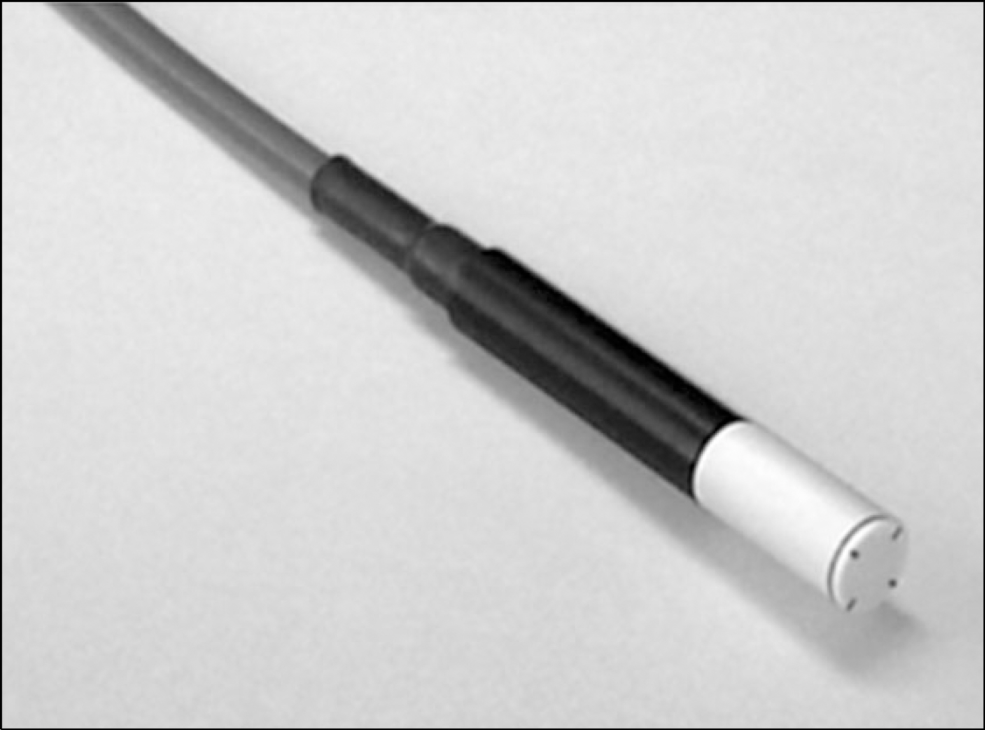
Fig. 1. Photo of the PTW 60019 microDiamond detector.
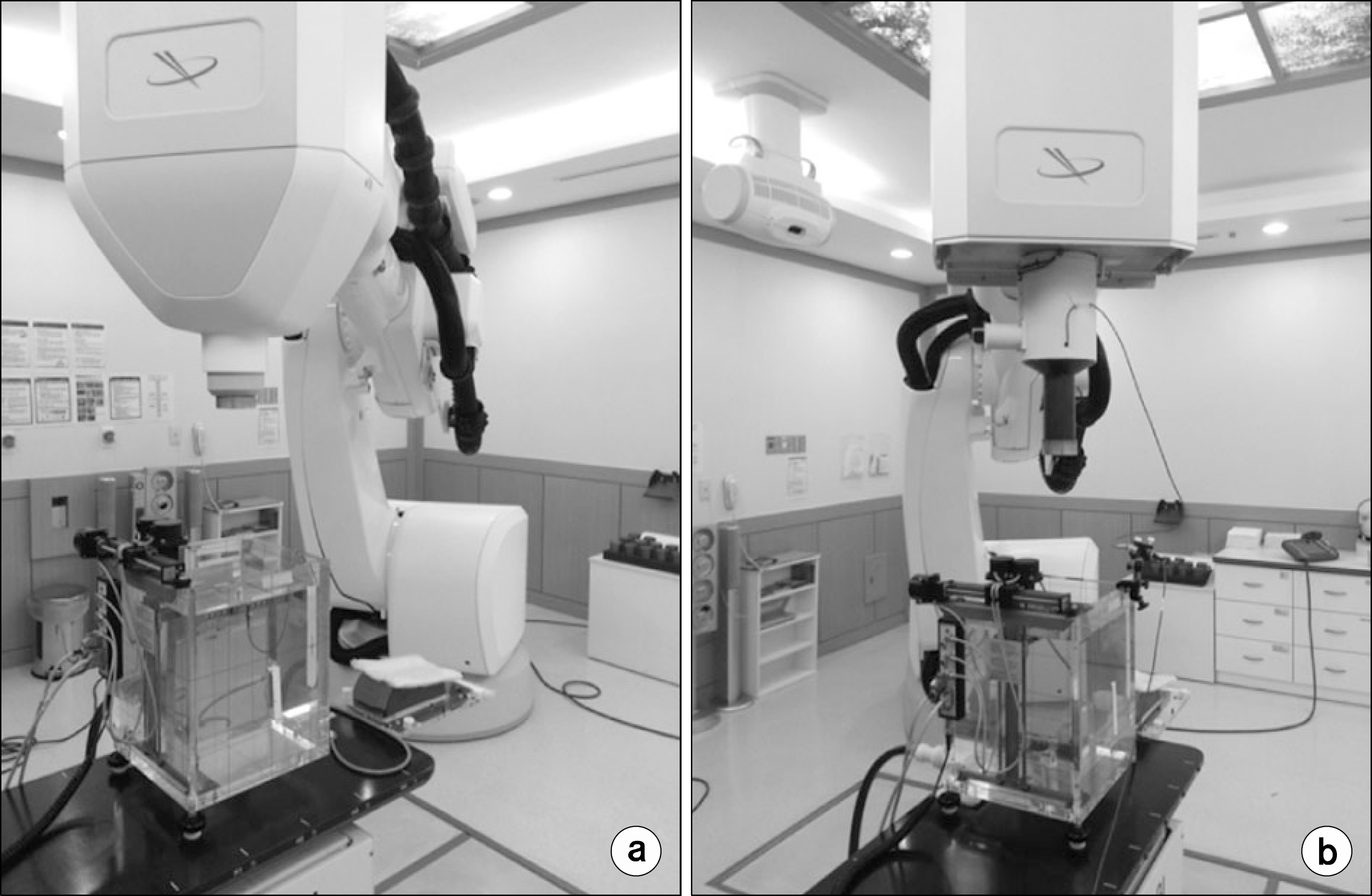
Fig. 2. Photographs depicting the experimental setup of cyberknife and water phantom. The source-detector distance of 80 cm at 1.5 g/cm2, 5 cm g/cm2 depth for measurement of (a) OF and (b) OCR, respectively. The measurement of dosimetic parameters with (b) PDD was scanned at SSD = 80 cm, from water surface to 25 cm depth.
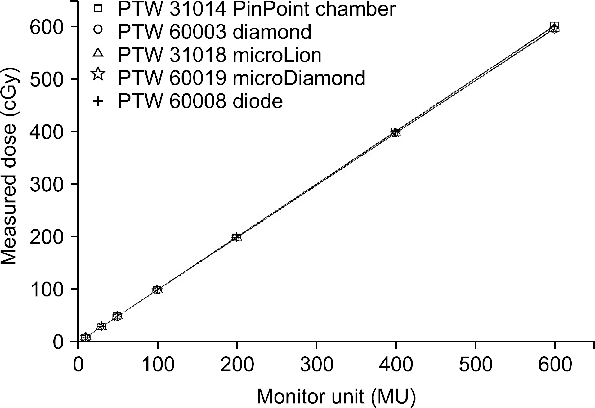
Fig. 3. The comparison of dose linearity for each detectors response were measured from 10 to 600 MU in 10×10 cm2 field with respect to the dose rate of 300 MU/min. Linear fit is plotted with solid line.
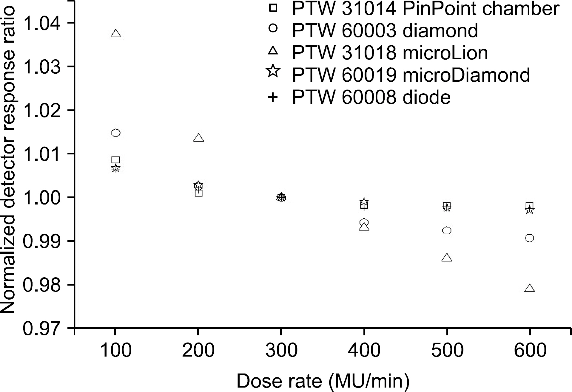
Fig. 4. The comparison of dose rate dependence for each detectors response were measured from 100 to 600 MU/min in 10×10 cm2 field at delivered dose 100 MU. Detector response at dose rate 300 MU/min was normalized to 1.

Fig. 5. The comparison of output factors for each detectors were measured in diameter from 0.5×0.5 cm2 to 6×6 cm2 with CyberKnife system.

Fig. 6. Depth-dose curves as measured with the each dectectors at (a) 5 mm, (b) 7.5 mm, (c) 10 mm, (d) 15 mm, (e) 20 mm, and (f) 30 mm collimator, respectively. All depth-dose curves are taken from water surface to 25 cm depth at SSD 100 cm.
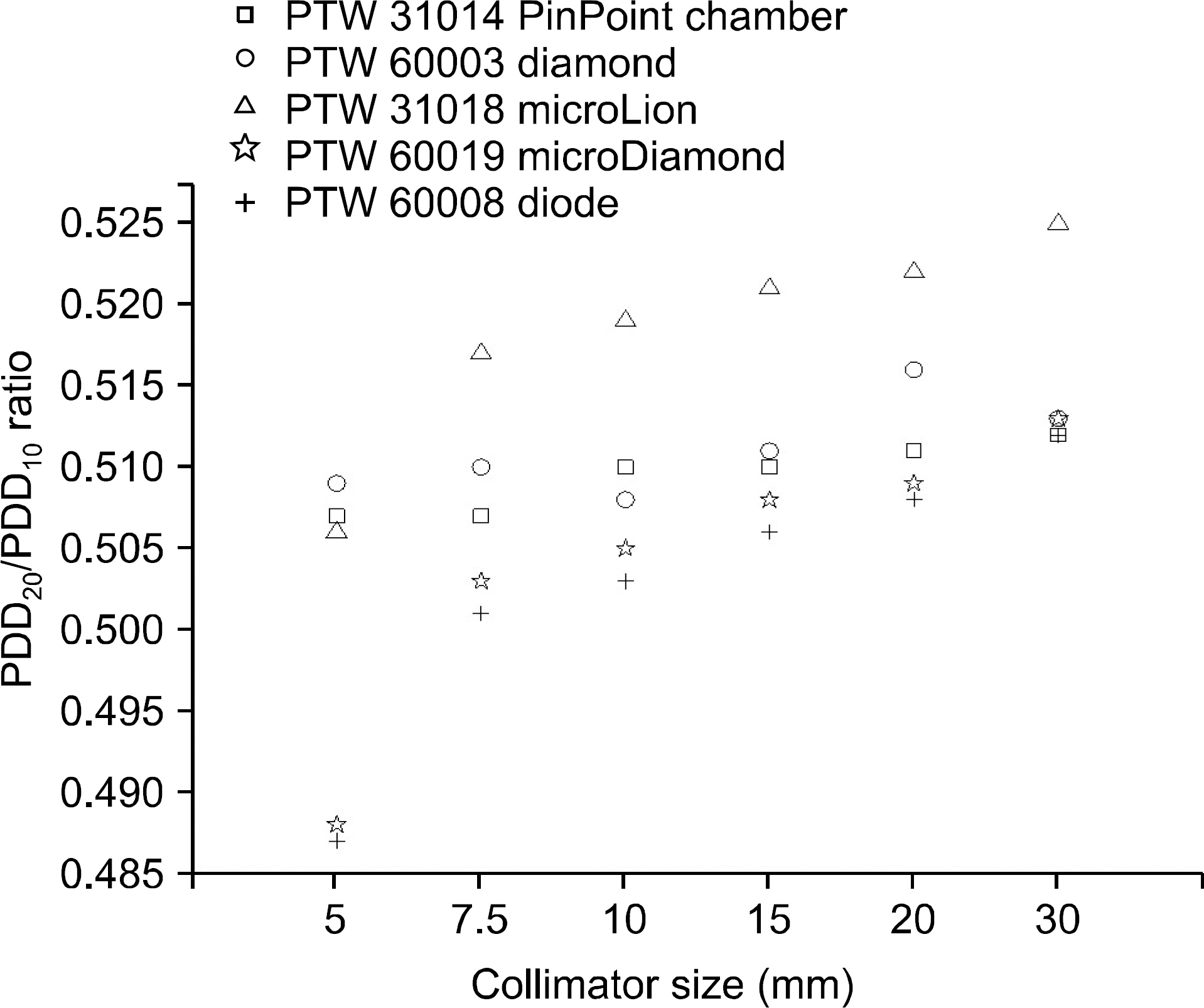
Fig. 7. PDD20/PDD10 ratio values derived from depth-dose curves.

Fig. 8. (a) The lateral dose profiles measured with the each detectors at 6 collimator size, respectively. All profiles are taken at 5 cm depth water at 80 cm SAD. (b) 80%∼20% penumbra values derived from the lateral beam profiles.
Reference
-
References
1. Wiggenraad RG, Petoukhova AL, Versluis L, et al. Stereotactic Radiotherapy of Intracranial Tumors: A Comparison of Intensity-Modulated Radiotherapy and Dynamic Conformal Arc. International Journal of Radiation Oncology Biology Physics. 74(4):1018–1026. 2009.
Article2. International Commission on Radiation Units and Measurements, Determination of absorbed dose in a patient irradiated by beams of X or Gamma rays in Radiotherapy procedure. ICRU Report No. 24 (. 1976.3. R. Alfonso, P. Andreo, R. Capote, et al: A new formalism for reference dosimetry of small and nonstandard fields. Med Phys. 35(11):5179–5186. 2008.4. I. J. Das, G. X. Ding, A. Ahnesjo: Small fields: Nonequilibrium radiation dosimetry. Med Phys. 35(1):206–215. 2007.5. C. McKerracher, D. I. Thwaites: Assessment of new small-field detectors against standard-field detectors for practical stereotactic beam data acquisition. Phys Med Biol. 44(9):2143–2160. 1999.6. Rice RK, Hansen JL, Svensson GK, et al. Measurements of Dose Distributions in Small Beams of 6 MV X-Rays. Physics in Medicine and Biology. 32(9):1087–1099. 1987.
Article7. Laub WU, Wong T. The Volume Effect of Detectors in the Dosimetry of Small Fields used in IMRT. The International Journal of Medical Physics Research and Practice. 30(3):341–347. 2003.
Article8. C. Bassinet, C. Huet, S. Derreumaux, et al: Small fields output factors measurements and correction factors determination for several detectors for a CyberKnifeR and linear accelerators equipped with microMLC and circular cones. Med Phys. 40(7):): 071725 (. 13. pp.) (. 2013.9. C. McKerracher, D. I. Thwaites: Head scatter ratios for small MV photon fields. Part II: The effects of source size and detector. Radiother Oncol. 85(2):286–291. 2007.10. P. H. Charles, G. Cranmer-Sargison, D. I. Thwaites et al: A practical and theoretical definition of very small field size for radiotherapy output factor measurements. Med Phys. 41(4):): 041707 (8pp.) (. 2014.11. P. Francescon, S. Cora, C. Cavedon: Total scatter factors of small beams: Amultidetector and Monte Carlo study. Med Phys. 35(2):504–513. 2008.12. H. Benmakhlouf, J. Sempau, P. Andreo: Output correction factors for nine small field detectors in 6MVradiation therapy photon beams: APENELOPE Monte Carlo study. Med Phys. 41(4):): 041711 (12pp.) (. 2014.13. P. Francescon, W. Kilby, N. Satariano: Monte Carlo simulated correction factors for output factor measurement with the CyberKnife system–Results for new detectors and correction factor dependence on measurement distance and detector orientation. Phys Med Biol. 59(6):): N11–17 (. 2014.14. Das IJ, Cheng CW, Watts RJ, et al. Accelerator Beam Data Commissioning Equipment and Procedures: Report of the TG-106 of the Therapy Physics Committee of the AAPM. The International Journal of Medical Physics Research and Practice. 35(9):4186–4215. 2008.
Article15. Sang Hyoun Choi, Hyun Do Huh, Seong Hoon Kim, et al. A Study of Characteristics of MicroLion Liquid Ionization Chamber for 6 MV Photon Beam, Progress in Medical Physics. 22(4):216–223. 2011.16. S. N. Rustgi, D. M. D. Frye: Dosimetric characterization of radiosurgical beams with a diamond detector. Med Phys. 22:2117–2121. 1995.17. P. W. Hoban, M. Heydarian, W. A. Beckham, et al: Dose rate dependence of a PTW diamond detector in the dosimetry of a 6 MV photon beam. Phys Med Biol. 39:1219–1229. 1994.18. G. Rikner: Silicon diode as detectors in relative dosimetry of photon, electron, and proton radiation fields. Ph.D. Thesis Uppsala University Sweden (. 1983.19. Sauer OA, Wilbert J. Measurement of output factors for small photon beams. Med Phys. 4:1983–1988. 2007.
Article20. Haryanto F, Fippel M, Laub W, et al. Investigation of Photon Beam Output Factors for Conformal Radiation Therapy-Monte Carlo Simulations and Measurements. Physics in Medicine and Biology. 47:N133–N143. 2002.21. Das IJ, Ding GX, Ahnesjo A. Small Fields: Nonequilibrium Radiation Dosimetry. The International Journal of Medical Physics Research and Practice. 35(1):206–215. 2008.
Article22. Francescon P, Cora S, Cavedon C. Total Scatter Factors of Small Beams: A Multidetector and Monte Carlo Study. The International Journal of Medical Physics Research and Practice. 35(2):504–513. 2008.
Article23. Kim JK, Wen N, Jin JY, et al. Clinical Commission and Use of the Novalis Tx Linear Accelerator for SRS and SBRT. Journal of Applied Clinical Medical Physics. 13(3):124–151. 2012.24. Johnny E Morales. Scott B. Crowe, Robin Hill, et al: Dosimetry of cone defined stereotatic radiosurgery fields with a commercial synthetic diamond detector. Med Phys. 41(11):111702. 2014.
- Full Text Links
-
- Actions
-
Cited
- CITED
-
- Close
- Share
-
- Similar articles
-
- Evaluation of the Dosimeter Volume Effect on Small-Field Dosimetry Using the Elekta Harmony Pro Linear Accelerator
- Dosimetric Characteristics of Edge Detector(TM) in Small Beam Dosimetry
- Feasibility of a Linear Diode Array Detector for Commissioning of a Radiotherapy Planning System
- Dosimetric Verifications of the Output Factors in the Small Field Less Than 3 cm2 Using the Gafchromic EBT2 Films and the Various Detectors
- Study for Multi Channel Radiation Detector Using of Microfilm and Carbon Electrode
