Prog Med Phys.
2023 Mar;34(1):1-9. 10.14316/pmp.2023.34.1.1.
Feasibility of a Linear Diode Array Detector for Commissioning of a Radiotherapy Planning System
- Affiliations
-
- 1Department of Radiation Oncology, Asan Medical Center, University of Ulsan College of Medicine, Seoul, Korea
- KMID: 2541141
- DOI: http://doi.org/10.14316/pmp.2023.34.1.1
Abstract
- Purpose
Although ionization chambers are widely used to measure beam commissioning data, point-by-point measurements of all the profiles with various field size and depths are timeconsuming tasks. As an alternative, we investigated the feasibility of a linear diode array for commissioning a treatment planning system.
Methods
The beam data of a Varian TrueBeam ® radiotherapy system at 6 and 10 MV with/without a flattening filter were measured for commissioning of an Eclipse Analytical Anisotropic Algorithm (AAA) ver.15.6. All of the necessary beam data were measured using an IBA CC13 ionization chamber and validated against Varian “Golden Beam” data. After validation, the measured CC13 profiles were used for commissioning the Eclipse AAA (AAA CC13 ). In addition, an IBA LDA-99SC linear diode array detector was used to measure all of the beam profiles and for commissioning a separate model (AAA LDA99 ). Finally, the AAA CC13 and AAA LDA99 dose calculations for each of the 10 clinical plans were compared.
Results
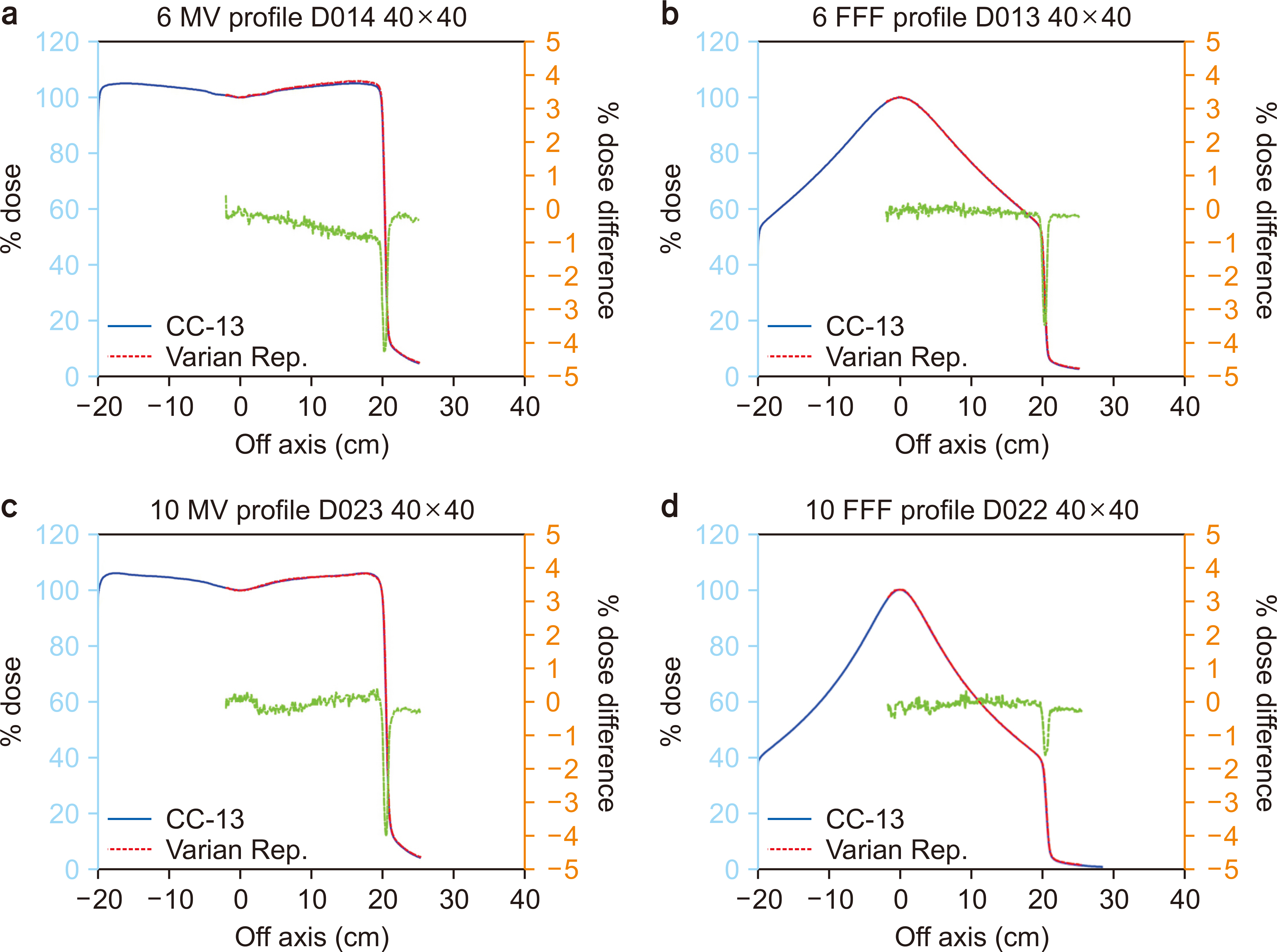
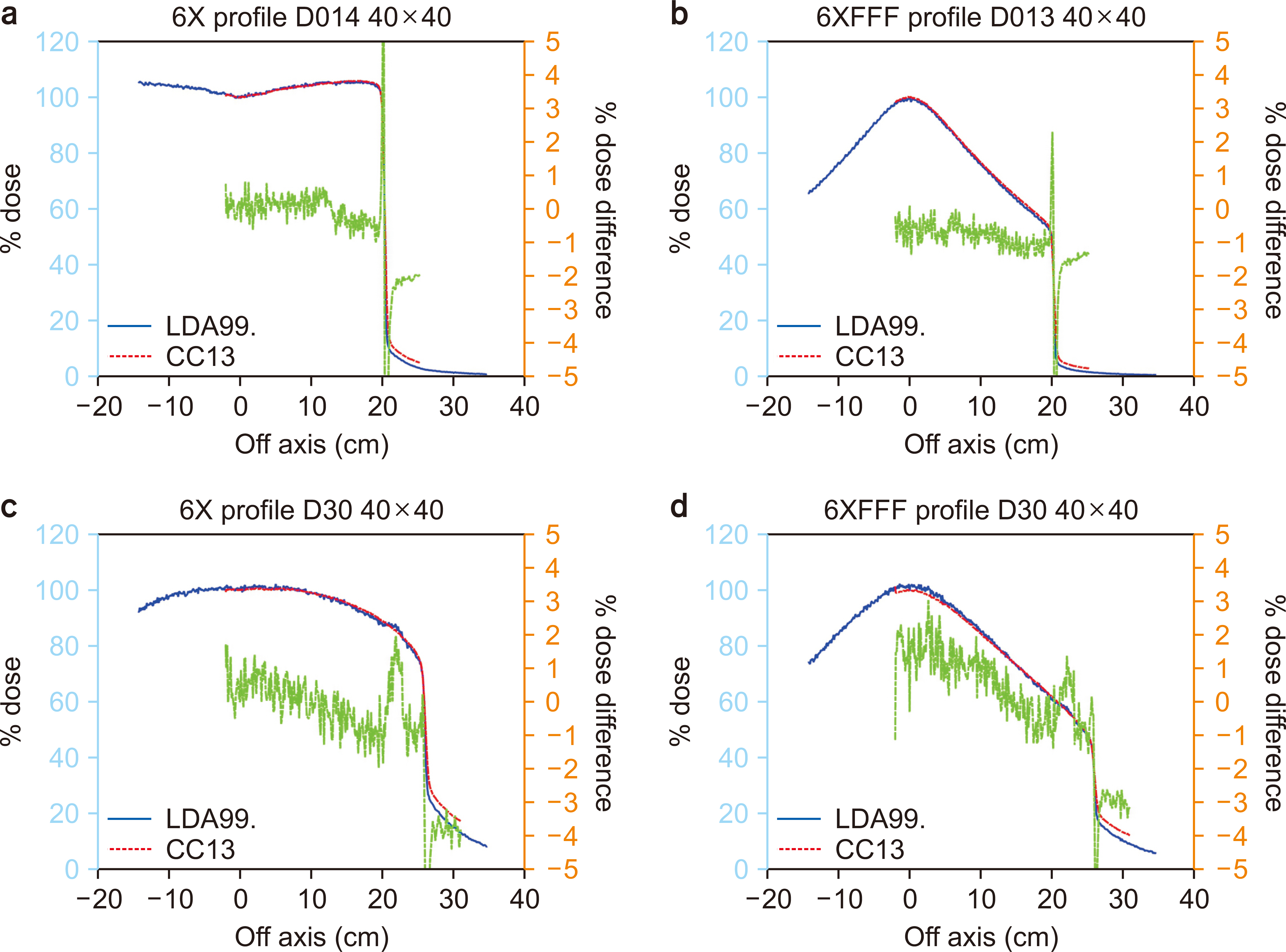
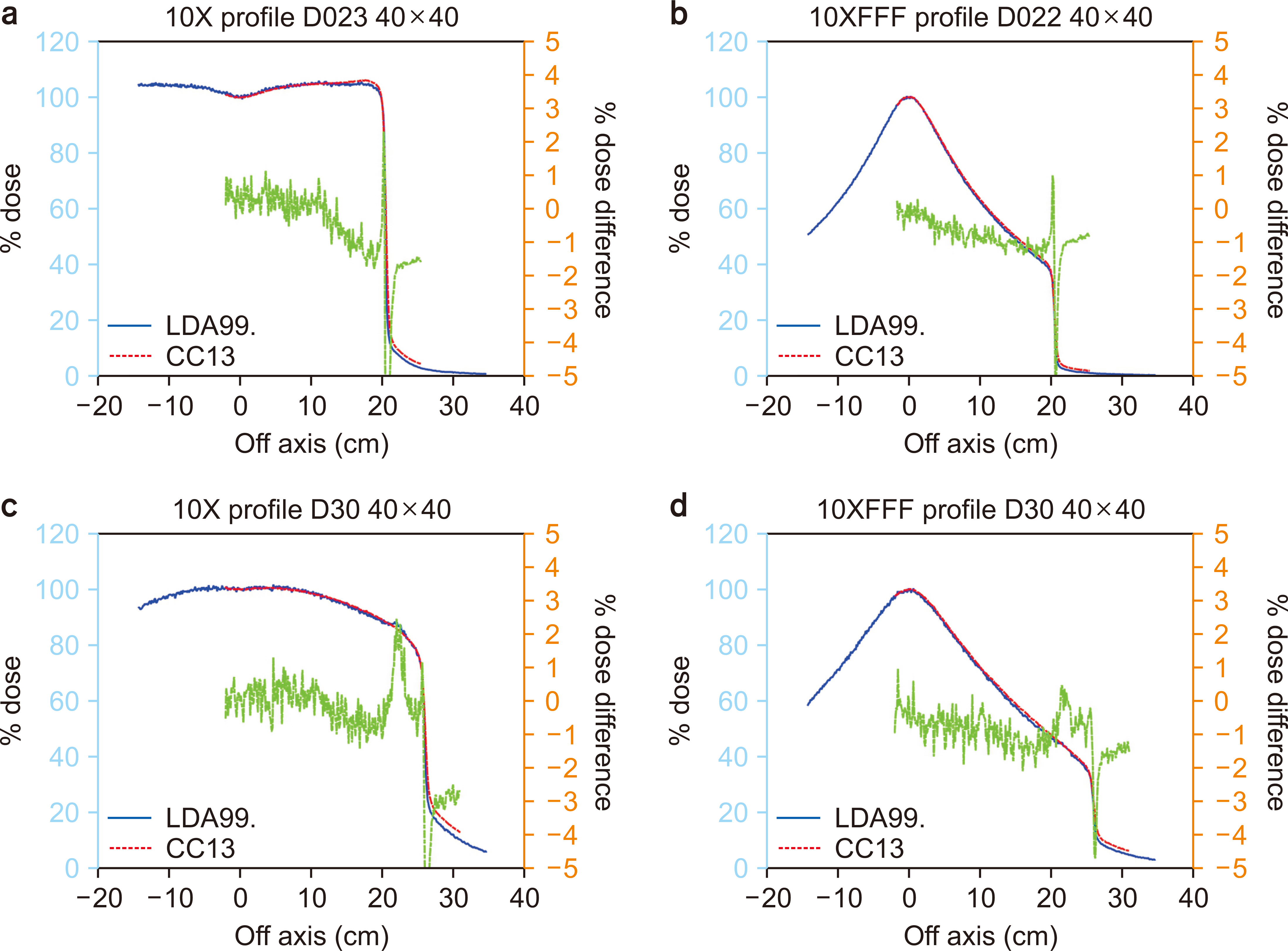
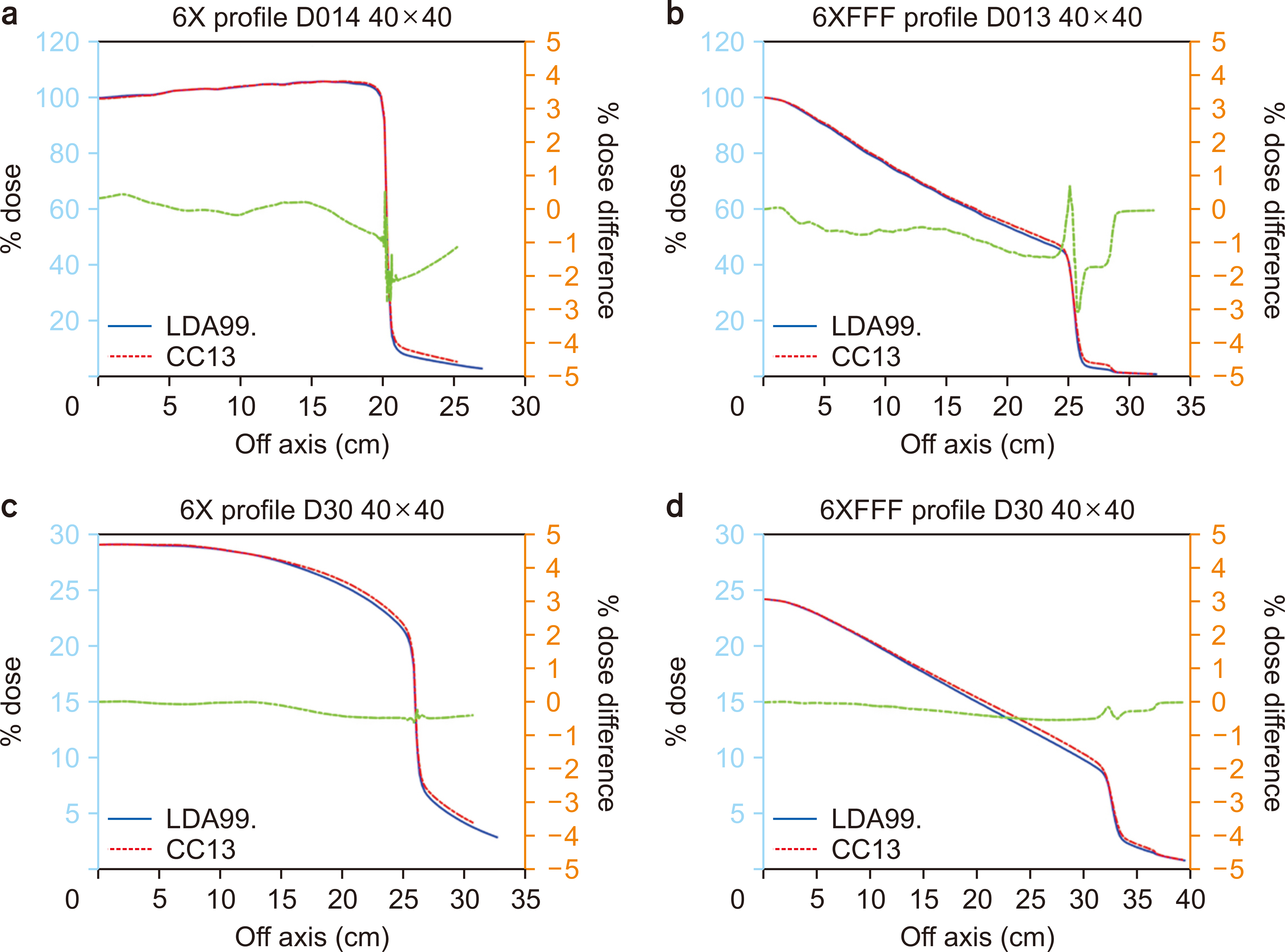
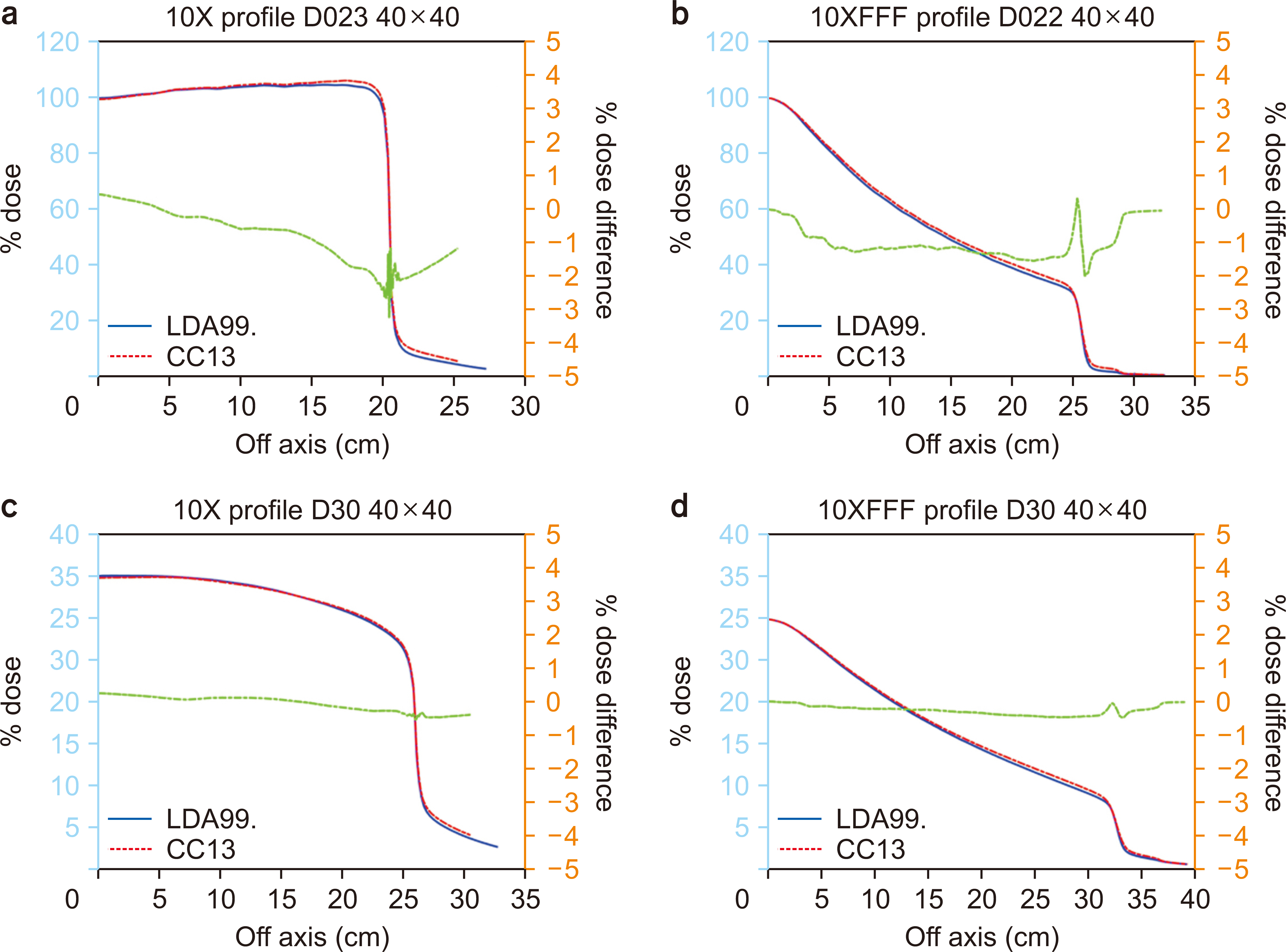
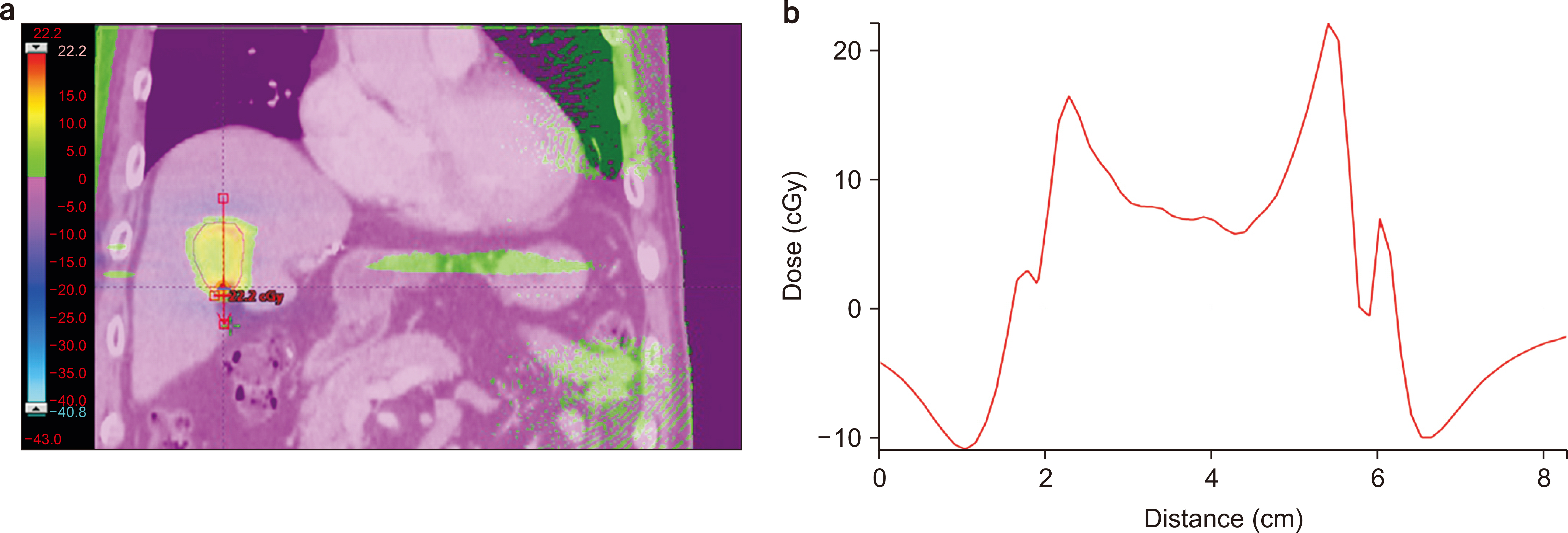
The agreement of the CC13 profiles with the Varian Golden Beam data was confirmed within 1% except in the penumbral region, where ≤2% of a discrepancy related to machinespecific jaw calibration was observed. Since the volume was larger for the CC13 chamber than for the LDA-99SC chamber, the penumbra widths were larger in the CC13 profiles, resulting in ≤5% differences. However, after beam modeling, the penumbral widths agreed within 0.1 mm. Finally the AAA LDA99 and AAA CC13 dose distributions agreed within 1% for all voxels inside the body for the 10 clinical plans.
Conclusions
In conclusion, the LDA-99SC diode array detector was found to be accurate and efficient for measuring photon beam profiles to commission treatment planning systems.
Keyword
Figure
Reference
-
References
1. Eklund K, Ahnesjö A. 2009; Modeling silicon diode energy response factors for use in therapeutic photon beams. Phys Med Biol. 54:6135–6150. DOI: 10.1088/0031-9155/54/20/007. PMID: 19779220.
Article2. Griessbach I, Lapp M, Bohsung J, Gademann G, Harder D. 2005; Dosimetric characteristics of a new unshielded silicon diode and its application in clinical photon and electron beams. Med Phys. 32:3750–3754. DOI: 10.1118/1.2124547. PMID: 16475774.
Article3. Glide-Hurst C, Bellon M, Foster R, Altunbas C, Speiser M, Altman M, et al. 2013; Commissioning of the Varian TrueBeam linear accelerator: a multi-institutional study. Med Phys. 40:031719. DOI: 10.1118/1.4790563. PMID: 23464314.
Article4. Palmans H, Andreo P, Huq MS, Seuntjens J, Christaki KE, Meghzifene A. 2018; Dosimetry of small static fields used in external photon beam radiotherapy: summary of TRS-483, the IAEA-AAPM international Code of Practice for reference and relative dose determination. Med Phys. 45:e1123–e1145. DOI: 10.1002/mp.13208. PMID: 30247757.
Article5. Barraclough B, Li JG, Lebron S, Fan Q, Liu C, Yan G. 2015; A novel convolution-based approach to address ionization chamber volume averaging effect in model-based treatment planning systems. Phys Med Biol. 60:6213–6226. DOI: 10.1088/0031-9155/60/16/6213. PMID: 26226323.
Article6. Fogliata A, Garcia R, Knoos T, Nicolini G, Clivio A, Vanetti E, et al. 2012; Definition of parameters for quality assurance of flattening filter free (FFF) photon beams in radiation therapy. Med Phys. 39:6455–6464. DOI: 10.1118/1.4754799. PMID: 23039680.
Article7. Wasbø E, Valen H. 2008; Dosimetric discrepancies caused by differing MLC parameters for dynamic IMRT. Phys Med Biol. 53:405–415. DOI: 10.1088/0031-9155/53/2/008. PMID: 18184995.
Article8. Eklund K, Ahnesjö A. 2010; Spectral perturbations from silicon diode detector encapsulation and shielding in photon fields. Med Phys. 37:6055–6060. DOI: 10.1118/1.3501316. PMID: 21158317.
Article
- Full Text Links
- Actions
-
Cited
- CITED
-
- Close
- Share
- Similar articles
-
- Analysis of Low MU Characteristics of Siemens Primus Linear Accelerator using Diode Arrays for IMRT QA
- Establishment of linear accelerator-based image guided radiotherapy for orthotopic 4T1 mouse mammary tumor model
- A Comparison Study of Volumetric Modulated Arc Therapy Quality Assurances Using Portal Dosimetry and MapCHECK 2
- Efficiency Study of 2D Diode Array Detector for IMRT Quality Assurance
- Proposal on Guideline for Quality Assurance of Radiation Treatment Planning System

