Pediatr Allergy Respir Dis.
2012 Sep;22(3):312-316.
A Case of Congenital Short Trachea Combined with Laryngeal Cleft
- Affiliations
-
- 1Department of Pediatrics, Samsung Medical Center, Sungkyunkwan University School of Medicine, Seoul, Korea. kmaped@skku.edu
- 2Environmental Health Center for Atopic Diseases, Samsung Medical Center, Sungkyunkwan University School of Medicine, Seoul, Korea.
- 3Department of Otorhinolaryngology-Head and Neck surgery, Samsung Medical Center, Sungkyunkwan University School of Medicine, Seoul, Korea.
Abstract
- Congenital short trachea is a rare congenital anomaly in which the trachea is composed of reduced number of cartilage rings, which result in an abnormally high position of the carina and an abnormal course of the main bronchi. Hazards of congenital short trachea in infants and children include inadvertent bronchial intubation, because it causes bronchiostenosis, pulmonary interstitial emphysema, pneumomediastinum, pneumothorax, and ipsilateral atelectasis. Laryngeal cleft is a rare condition, as well. Symptoms range from mild stridor to massive aspiration and respiratory distress, depending on the severity of the cleft. Until now, a case with combination of these two rare congenital defects has not been reported. Herein, we report a 13 month-old boy who has congenital short trachea with laryngeal cleft.
Keyword
MeSH Terms
Figure
-
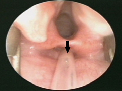
Fig. 1 Type 2 laryngeal cleft in laryngoscopy (arrow).
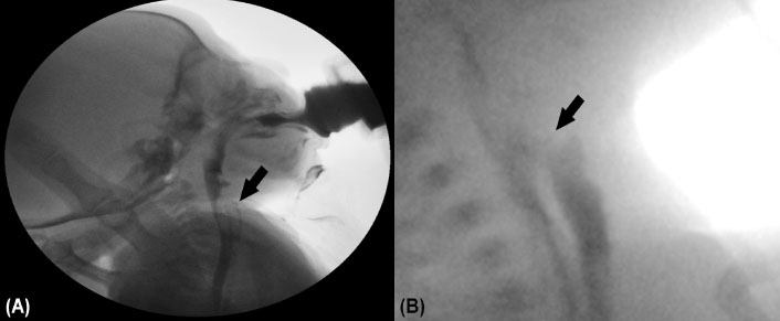
Fig. 2 (A) Video fluoroscopic swallowing study and (B) esophagography show contrast is leaking between larynx and esophagus.
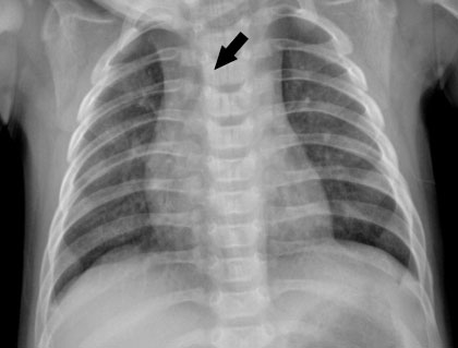
Fig. 3 Chest X-ray demonstrating carina at the level of T3 (arrow).
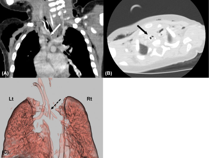
Fig. 4 (A, B) Trachea computed tomography image reveals carina at the level of T3 and endotracheal tube in the right bronchus (solid line arrow). (C) Trachea computed tomography 3D reconstruction image reveals diffuse narrowing of right main bronchus (dot line arrow). Lt, left; Rt, right.
Reference
-
1. Wells AL, Wells TR, Landing BH, Cruz B, Galvis DA. Short trachea, a hazard in tracheal intubation of neonates and infants: syndromal associations. Anesthesiology. 1989. 71:367–373.2. van der Doef HP, Yntema JB, van den Hoogen FJ, Marres HA. Clinical aspects of type 1 posterior laryngeal clefts: literature review and a report of 31 patients. Laryngoscope. 2007. 117:859–863.
Article3. de Priester JA, Vos GD, van Waardenburg DA, Oei TK. Congenitally short trachea with compression of the left mainstem bronchus: MRI findings. Pediatr Radiol. 1998. 28:342–343.
Article4. Ravenna F, Caramori G, Panella GL, Papi A, Benea G, Adcock IM, et al. An unusual case of congenital short trachea with very long bronchi mimicking bronchial asthma. Thorax. 2002. 57:372–373.
Article5. Rahbar R, Rouillon I, Roger G, Lin A, Nuss RC, Denoyelle F, et al. The presentation and management of laryngeal cleft: a 10-year experience. Arch Otolaryngol Head Neck Surg. 2006. 132:1335–1341.6. Frazer JE. The development of the larynx. J Anat Physiol. 1910. 44(Pt 2):156–191.7. Pezzettigotta SM, Leboulanger N, Roger G, Denoyelle F, Garabédian EN. Laryngeal cleft. Otolaryngol Clin North Am. 2008. 41:913–933. ix
Article8. Thiel G, Clement WA, Kubba H. The management of laryngeal clefts. Int J Pediatr Otorhinolaryngol. 2011. 75:1525–1528.
Article9. Wells TR, Jacobs RA, Senac MO, Landing BH. Incidence of short trachea in patients with myelomeningocele. Pediatr Neurol. 1990. 6:109–111.
Article10. Wells TR, Wells AL, Galvis DA, Senac MO Jr, Landing BH, Vachon LA. Diagnostic aspects and syndromal associations of short trachea with bronchial intubation. Am J Dis Child. 1990. 144:1369–1371.
Article11. Leboulanger N, Garabedian EN. Laryngo-tracheo-oesophageal clefts. Orphanet J Rare Dis. 2011. 6:81.
Article12. Hu CH, Liu YF, Yu JS, Ng YY, Chen SJ, Su PH, et al. A MID1 gene mutation in a patient with Opitz G/BBB syndrome that altered the 3D structure of SPRY domain. Am J Med Genet A. 2012. 158A:726–731.
Article13. Chien W, Ashland J, Haver K, Hardy SC, Curren P, Hartnick CJ. Type 1 laryngeal cleft: establishing a functional diagnostic and management algorithm. Int J Pediatr Otorhinolaryngol. 2006. 70:2073–2079.
Article
- Full Text Links
-
- Actions
-
Cited
- CITED
-
- Close
- Share
-
- Similar articles
-
- A case of laryngeal cleft
- A Case of CongenitaI LaryngeaI Cleft
- A Case of Congenital Deformity of the Eye, Combined with Cleft Lip and Palate
- Unanticipated Difficult Intubation in a Patient with Asymptomatic Congenital Subglottic Stenosis Caused by a Laryngeal Web and Thyroid Cartilage Anomaly: A case report
- Combined Congenital Anterior and Posterior Midline Cleft of the Atlas Associated with Asymptomatic Lateral Atlantoaxial Subluxation
