Lab Med Online.
2012 Apr;2(2):95-100.
Relationship of White-Matter Lesions and Lacunar Infarcts with Cardiovascular Risk Factors
- Affiliations
-
- 1Korea Association of Health Promotion, Seoul, Korea. cellonah@hanmail.net
Abstract
- BACKGROUND
Magnetic resonance imaging (MRI) findings of white-matter lesions are different from those of lacunar infarcts; however, both these conditions are related to cardiovascular risk factors. This study was performed to investigate the differences in the relationships of white-matter lesions and lacunar infarcts with cardiovascular risk factors and differences between the metabolic characteristics of patients with these conditions.
METHODS
We included 4,255 patients who showed neurological deficits during health checkups. These individuals were classified into the following 3 groups on the basis of MRI findings: normal, white-matter lesion, and lacunar infarct. The groups were compared for age; weights; prevalence of metabolic syndrome; and levels of blood pressure, blood glucose, lipid, high sensitivity C-reactive protein, and HbA1c.
RESULTS
Age, body mass index (BMI); waist circumference; levels of blood pressure, blood glucose, triglycerides and HbA1c; and prevalence of metabolic syndrome and its components were the highest in lacunar infarct group, followed by white matter lesion group, and normal group. Age and diastolic blood pressure level were related to white matter lesions, and age, systolic blood pressure level, and blood glucose level were related to lacunar infarcts. Further, the prevalence of the above-mentioned lesions increased with increase of the number of the components of metabolic syndrome.
CONCLUSIONS
This study suggests that lacunar infarct is more advanced lesion than white matter lesion. Among all the cardiovascular risk factors, high blood pressure and impaired fasting blood glucose levels were significantly related to white-matter lesions and lacunar infarct.
MeSH Terms
Figure
-
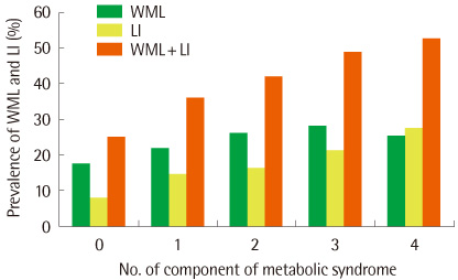
Fig. 1 Prevalence of white-matter lesions and lacunar infarct according to the number of the components of metabolic syndrome. P<0.01 derived from χ2 tests used to comparison the prevalence of white-matter lesion and lacunar infarct with reference to the number of components of the metabolic syndrome. Abbreviations: WML, white-matter lesion; LI, lacunar infarct.
Reference
-
1. Vermeer SE, Prins ND, den Heijer T, Hofman A, Koudstaal PJ, Breteler MM. Silent brain infarcts and the risk of dementia and cognitive decline. N Engl J Med. 2003. 348:1215–1222.
Article2. Vermeer SE, Hollander M, van Dijk EJ, Hofman A, Koudstaal PJ, Breteler MM. Silent brain infarcts and white matter lesions increase stroke risk in the general population: the Rotterdam Scan Study. Stroke. 2003. 34:1126–1129.
Article3. Vermeer SE, Longstreth WT Jr, Koudstaal PJ. Silent brain infarcts: a systematic review. Lancet Neurol. 2007. 6:611–619.
Article4. Kobayashi S, Okada K, Koide H, Bokura H, Yamaguchi S. Subcortical silent brain infarction as a risk factor for clinical stroke. Stroke. 1997. 28:1932–1939.
Article5. Pantoni L, Garcia JH. Pathogenesis of leukoaraiosis: a review. Stroke. 1997. 28:652–659.6. de Leeuw FE, de Groot JC, Oudkerk M, Witteman JC, Hofman A, van Gijn J, et al. A follow-up study of blood pressure and cerebral white matter lesions. Ann Neurol. 1999. 46:827–833.
Article7. Wardlaw JM, Sandercock PA, Dennis MS, Starr J. Is breakdown of the blood-brain barrier responsible for lacunar stroke, leukoaraiosis, and dementia. Stroke. 2003. 34:806–812.
Article8. Bamford JM, Warlow CP. Evolution and testing of the lacunar hypothesis. Stroke. 1988. 19:1074–1082.
Article9. Fernando MS, Simpson JE, Mathews F, Brayne C, Lewis CE, Barber R, et al. White matter lesions in an unselected cohort of the elderly: molecular pathology suggests origin from chronic hypoperfusion injury. Stroke. 2006. 37:1391–1398.
Article10. Gállego J, Martínez-Vila E. Asymptomatic cerebrovascular disease and systemic diagnosis in stroke, atherothrombosis as a disease of the vascular tree. Cerebrovasc Dis. 2005. 20:Suppl 2. 1–10.
Article11. Launer LJ. Epidemiology of white matter lesions. Top Magn Reson Imaging. 2004. 15:365–367.
Article12. Grundy SM, Cleeman JI, Daniels SR, Donato KA, Eckel RH, Franklin BA, et al. Diagnosis and management of the metabolic syndrome: an American Heart Association/National Heart, Lung, and Blood Institute Scientific Statement. Circulation. 2005. 112:2735–2752.13. WHO West Pacific Region. The Asia-pacific perspective: refining obesity and its treatment. 2000. London: International Obesity Taskforce.14. Prins ND, van Dijk EJ, den Heijer T, Vermeer SE, Jolles J, Koudstaal PJ, et al. Cerebral small-vessel disease and decline in information processing speed, executive function and memory. Brain. 2005. 128:2034–2041.
Article15. Sonohara K, Kozaki K, Akishita M, Nagai K, Hasegawa H, Kuzuya M, et al. White matter lesions as a feature of cognitive impairment, low vitality and other symptoms of geriatric syndrome in the elderly. Geriatr Gerontol Int. 2008. 8:93–100.
Article16. Bernick C, Kuller L, Dulberg C, Longstreth WT Jr, Manolio T, Beauchamp N, et al. Silent MRI infarcts and the risk of future stroke: the cardiovascular health study. Neurology. 2001. 57:1222–1229.
Article17. Pantoni L. Cerebral small vessel disease: from pathogenesis and clinical characteristics to therapeutic challenges. Lancet Neurol. 2010. 9:689–701.
Article18. Atwood LD, Wolf PA, Heard-Costa NL, Massaro JM, Beiser A, D'Agostino RB, et al. Genetic variation in white matter hyperintensity volume in the Framingham Study. Stroke. 2004. 35:1609–1613.
Article19. McNeill AM, Rosamond WD, Girman CJ, Golden SH, Schmidt MI, East HE, et al. The metabolic syndrome and 11-year risk of incident cardiovascular disease in the atherosclerosis risk in communities study. Diabetes Care. 2005. 28:385–390.
Article20. Klein BE, Klein R, Lee KE. Components of the metabolic syndrome and risk of cardiovascular disease and diabetes in Beaver Dam. Diabetes Care. 2002. 25:1790–1794.
Article21. Kwon HM, Kim BJ, Lee SH, Choi SH, Oh BH, Yoon BW. Metabolic syndrome as an independent risk factor of silent brain infarction in healthy people. Stroke. 2006. 37:466–470.
Article22. American Psychiatric Association. Diagnostic and statistical manual of mental disorders. 1994. 4th ed. Washington: American Psychiatric Association.23. Manolio TA, Furberg CD, Shemanski L, Psaty BM, O'Leary DH, Tracy RP, et al. The CHS Collaborative Research Group. Associations of postmenopausal estrogen use with cardiovascular disease and its risk factors in older women. Circulation. 1993. 88:2163–2171.
Article24. Das RR, Seshadri S, Beiser AS, Kelly-Hayes M, Au R, Himali JJ, et al. Prevalence and correlates of silent cerebral infarcts in the Framingham offspring study. Stroke. 2008. 39:2929–2935.
Article25. Vermeer SE, Koudstaal PJ, Oudkerk M, Hofman A, Breteler MM. Prevalence and risk factors of silent brain infarcts in the population-based Rotterdam scan study. Stroke. 2002. 33:21–25.
Article26. Khan U, Porteous L, Hassan A, Markus HS. Risk factor profile of cerebral small vessel disease and its subtypes. J Neurol Neurosurg Psychiatry. 2007. 78:702–706.
Article27. Park K, Yasuda N, Toyonaga S, Tsubosaki E, Nakabayashi H, Shimizu K. Significant associations of metabolic syndrome and its components with silent lacunar infarction in middle aged subjects. J Neurol Neurosurg Psychiatry. 2008. 79:719–721.
Article28. Longstreth WT Jr, Manolio TA, Amold A, Burke GL, Bryan N, Jungreis CA, et al. Clinical correlates of white matter findings on cranial magnetic resonance imaging of 3301 elderly people. The Cardiovascular Health Study. Stroke. 1996. 27:1274–1282.
Article29. Dufouil C, de Kersaint-Gilly A, Besancon V, Levy C, Auffray E, Brunnereau L, et al. Longitudinal study of blood pressure and white matter hyperintensities: the EVA MRI Cohort. Neurology. 2001. 56:921–926.
Article
- Full Text Links
-
- Actions
-
Cited
- CITED
-
- Close
- Share
-
- Similar articles
-
- Is Obstructive Sleep Apnea a Risk Factor of Subclinical White Matter Damages?
- The Correlation of the White Matter Lesions and Lacunar Infarcts in Patients with Vascular Cognitive Impairment
- Clinical Analysis for Risk Factors of Lacunar Infarction Syndrome
- Evolving Concept of Small Vessel Disease through Advanced Brain Imaging
- Brain MRI Findings of Carbon Disulfide Poisoning
