Lab Anim Res.
2010 Jun;26(2):211-213. 10.5625/lar.2010.26.2.211.
Uterine Adenomyosis in Beagle Dogs
- Affiliations
-
- 1College of Veterinary Medicine & Institute of Veterinary Science, Kangwon National University, Chuncheon, Korea. byoon@kangwon.ac.kr
- 2Preclinical Research Center, Chemon Inc., Yongin, Korea.
- 3College of Veterinary Medicine, Jeju National University, Jeju, Korea.
- 4Department of Biomedical Laboratory Science, Namseoul University, Chonan, Korea.
- 5Peer Review Working Group of Preclinical Research Center, Chemon Inc., Yongin, Korea.
- KMID: 2312067
- DOI: http://doi.org/10.5625/lar.2010.26.2.211
Abstract
- Adenomyosis is a nonneoplastic hyperplastic lesion, characterized by invagination of proliferating endometrial glands into myometrium. In dogs, uterine adenomyosis is relatively rare and it is important in Toxicologic Pathology to differentiate other non-neoplastic and neoplastic lesions in uterus. In the present study, we report two cases of adenomyosis in the female beagle dogs used for a chemical toxicity test. Clinically, one out of the two female beagle dogs, 15 months of age, had vaginal bleeding for 2 weeks and the other one, 11 months of age, showed swelling of vulva for a week. At necropsy, the weight of uterus was markedly increased to 27.9 g and 15.8 g, compared with the mean value (4.01+/-2.37, n=6) of that of other normal dogs, respectively. The parameters of hematology and serum chemistry were ranged normal in both of the dogs with enlarged uterus. For differentiation of connective tissue with muscle fibers, Van Gieson stain was also performed in the serial tissue sections. Histopathologically, the lesions of the enlarged uteruses were characterized by proliferating endometrial glands into myometrium, surrounded by connective tissue. The endometrial glands were proliferating downward to myometrium or embedded in multiple clustered glands in deeper myometrium without compressing the adjacent muscle fibers. The gland epithelial cells are uniformly cuboidal shape with a dense and bottom-located nucleus. These gross and histological findings were consistent with adenomyosis.
Keyword
MeSH Terms
Figure
-
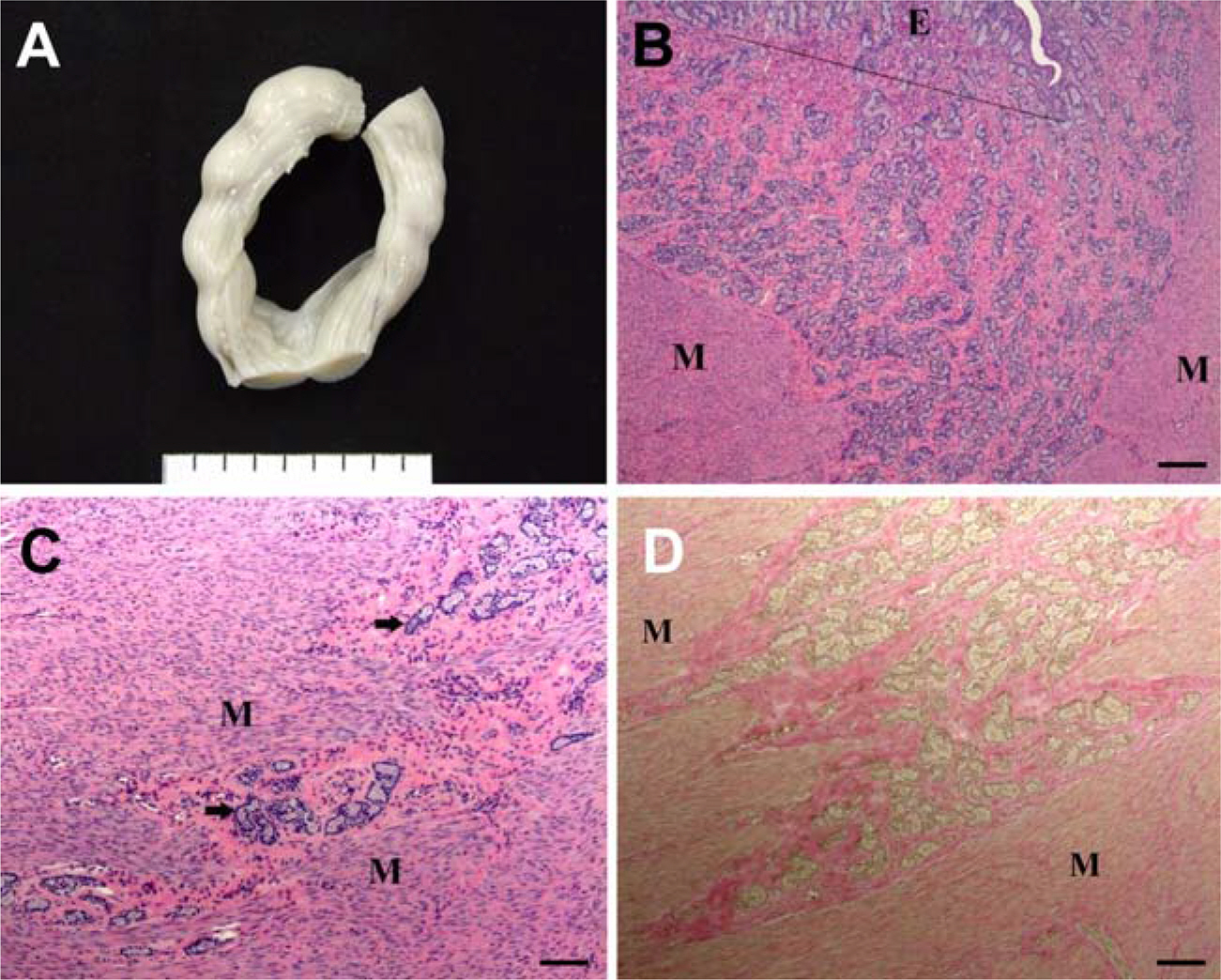
Figure 1. Gross and microscopic findings of adenomyosis. The uteri of the dogs were markedly enlarged (A). Note the proliferating endometrial glands into myometrium (M) from endometrium (E, upper of line) (B). Some multiple clustered endometrial glands were also found with being embedded in deeper myometrium without compressing the adjacent muscle fibers (C). The glandular epithelial cells are uniformly cuboidal shape without cellular atypia. The glands were surrounded by connective tissue (deep red in D). H&E stain for B and C and Van Gieson stain for D. Bars=100 µm for C and D and 200 µm for B.
Reference
-
Bergeron C.., Amant F.., Ferency A.2006. Pathology and physiopathology of adenomyosis. Best Pract. Res. Clin. Obstet. Gynaecol. 20:511–521.
ArticleBulman-Fleming J.2008. A rare case of uterine adenomyosis in a Siamese cat. Can. Vet. J. 49:709–712.Graham K.J.., Hulst F.A.., Vogelnest L.., Fraser I.S.., Shilton C.M.2009. Uterine adenomyosis in an organ-utan (Pongo abelii/pygmaeus). Aust. Vet. J. 87:66–69.Greaves P.1990. Histopathology of Preclinical Toxicity Studies: Interpretation and Relevance in Drug Safety Evaluation. 1st ed., pp. 638-639, Elsevier, Amsterdam.Haschek W.M.., Rousseaux C.G.1998. Fundamentals of Toxicologic Pathology. 1st ed., pp. 485-514, Academic Press, San Diego.Hur H.M.., Jung J.Y.., Kang S.C.., Park D.S.., Bae J.H.., Kim J.H.2008. Uterine adenomyosis in a cat. J. Vet. Clin. 25:19–22.Leininger J.R.., Jokinen M.P.1990. Pathology of the Fischer Rat. 1st ed., pp. 443-459, Academic Press, London.MacLachlan N.J.., Kennedy P.C.2002. Tumors in Domestic Animals. 4th ed., pp. 547-573, Blackwell Publishing, Ames.Nielsen S.W.., Kennedy P.C.1990. Tumors in Domestic Animals. 3rd ed., pp. 479-517, University of California Press, London.Otsuki Y.., Misaki O.., Sugimoto O., et al. 1994. Cyclic bcl-2 gene expression in human uterine endometrium during menstrual cycle. Lancet. 344:28–29.
ArticleSchlafer D.H.., Gifford A.T.2008. Cystic endometrial hyperplasia, pseudo-plancentational endometrial hyperplasia, and other cystic conditions of the canine and feline uterus. Theriogenology. 70:349–358.Tamada H.., Kawate N.., Inaba T.., Kuwamura M.., maeda M.., Kajikawa T.., Sawada T.2005. Adenomyosis with severe inflammation in the uterine cervix in a dog. Can. Vet. J. 46:333–334.
- Full Text Links
-
- Actions
-
Cited
- CITED
-
- Close
- Share
-
- Similar articles
-
- Recurrent Cerebral Infarction Associated with Uterine Adenomyosis
- Current status of high-intensity focused ultrasound for the management of uterine adenomyosis
- Are Transvaginal Sonography and Color doppler findings Useful for the Diagnosis of Adenomyosis?
- Preterm delivery in a primigravida with uterine adenomyosis
- Uterine myoma and adenomyosis: sonographic findings and differentiation
