Anat Cell Biol.
2016 Jun;49(2):99-106. 10.5115/acb.2016.49.2.99.
Changes in expression of proteasome in rats at different stages of atherosclerosis
- Affiliations
-
- 1Department of Biochemistry, Faculty of Medicine, Riau University, Pekanbaru, Indonesia. ismawati75@yahoo.com
- 2Department of Biochemistry, Faculty of Medicine, Andalas University, Padang, Indonesia.
- 3Department of Anatomy, Faculty of Medicine, Andalas University, Padang, Indonesia.
- KMID: 2308916
- DOI: http://doi.org/10.5115/acb.2016.49.2.99
Abstract
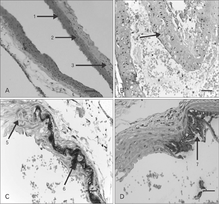
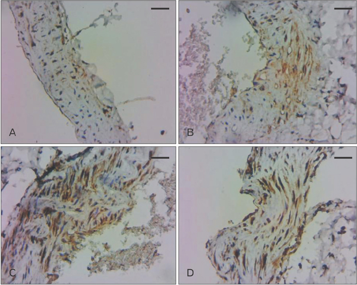
- It has been suggested that proteasome system has a role in initiation, progression, and complication stages of atherosclerosis. Although there is still controversy, there has been no research that compares the expression of proteasome in tissue and serum at each of these stages. This study aimed to investigated the expression of proteasome at different stages of atherosclerosis using rat model. We measured the expression of aortic proteasome by immunohistochemical analyses and were then analyzed using ImageJ software for percentage of area and integrated density. We used Photoshop version 3.0 to analyze aortic proteasome expression as a comparison. We measured serum proteasome expression by enzyme linked immunosorbents assays. Kruskal-Wallis test was used to compare mean value of percentage of area and serum proteasome. Analysis of variance test was used to compare mean value of integrated density. Correlation test between vascular proteasome expression and serum proteasome expression was made using Spearman test. A P-value of 0.05 was considered statistically significant. Compared with normal, percentage of area was higher in initiation, progression, and complication. Compared with normal, integrated density was higher in initiation and further higher in progression and complication. Data from Image J is similar with data from Photoshop. Serum proteasome expression was higher in initiation compared with normal, and further higher in progression and complication. It was concluded that there were different vascular proteasome expression and serum proteasome expression at the stages of atherosclerosis. These results may be used in research into new marker and therapeutic target in atherosclerosis.
Keyword
MeSH Terms
Figure
Cited by 1 articles
-
The effect of alpha-lipoic acid on expression of VCAM-1 in type 2 diabetic rat
Ismawati, Mukhyarjon, Enikarmila Asni, Ilhami Romus
Anat Cell Biol. 2019;52(2):176-182. doi: 10.5115/acb.2019.52.2.176.
Reference
-
1. World Health Organization. Cardiovascular diseases fact sheet [Internet]. Geneva: World Health Organization;2009. cited 2009 Aug 17. Available from: http://www.who.int/mediacentre/factsheets/fs317/en/print.html.2. Lloyd-Jones D, Adams R, Carnethon M, De Simone G, Ferguson TB, Flegal K, Ford E, Furie K, Go A, Greenlund K, Haase N, Hailpern S, Ho M, Howard V, Kissela B, Kittner S, Lackland D, Lisabeth L, Marelli A, McDermott M, Meigs J, Mozaffarian D, Nichol G, O'Donnell C, Roger V, Rosamond W, Sacco R, Sorlie P, Stafford R, Steinberger J, Thom T, Wasserthiel-Smoller S, Wong N, Wylie-Rosett J, Hong Y. American Heart Association Statistics Committee and Stroke Statistics Subcommittee. Heart disease and stroke statistics: 2009 update: a report from the American Heart Association Statistics Committee and Stroke Statistics Subcommittee. Circulation. 2009; 119:e21–e181.3. Pyle AL, Young PP. Atheromas feel the pressure: biomechanical stress and atherosclerosis. Am J Pathol. 2010; 177:4–9.4. Stocker R, Keaney JF Jr. Role of oxidative modifications in atherosclerosis. Physiol Rev. 2004; 84:1381–1478.5. Stary HC, Chandler AB, Dinsmore RE, Fuster V, Glagov S, Insull W Jr, Rosenfeld ME, Schwartz CJ, Wagner WD, Wissler RW. A definition of advanced types of atherosclerotic lesions and a histological classification of atherosclerosis. A report from the Committee on Vascular Lesions of the Council on Arteriosclerosis, American Heart Association. Circulation. 1995; 92:1355–1374.6. Fonarow GC. Aggressive treatment of atherosclerosis: the time is now. Cleve Clin J Med. 2003; 70:431–434. 437–438. 4407. Herrmann J, Lerman LO, Lerman A. On to the road to degradation: atherosclerosis and the proteasome. Cardiovasc Res. 2010; 85:291–302.8. Herrmann J, Ciechanover A, Lerman LO, Lerman A. The ubiquitin-proteasome system in cardiovascular diseases: a hypothesis extended. Cardiovasc Res. 2004; 61:11–21.9. Sixt SU, Beiderlinden M, Jennissen HP, Peters J. Extracellular proteasome in the human alveolar space: a new housekeeping enzyme? Am J Physiol Lung Cell Mol Physiol. 2007; 292:L1280–L1288.10. Marfella R, D'Amico M, Di Filippo C, Siniscalchi M, Sasso FC, Ferraraccio F, Rossi F, Paolisso G. The possible role of the ubiquitin proteasome system in the development of atherosclerosis in diabetes. Cardiovasc Diabetol. 2007; 6:35.11. Russell JC, Proctor SD. Small animal models of cardiovascular disease: tools for the study of the roles of metabolic syndrome, dyslipidemia, and atherosclerosis. Cardiovasc Pathol. 2006; 15:318–330.12. Li J, Chen CX, Shen YH. Effects of total glucosides from paeony (Paeonia lactiflora Pall) roots on experimental atherosclerosis in rats. J Ethnopharmacol. 2011; 135:469–475.13. Vinitha R, Thangaraju M, Sachdanandam P. Effect of tamoxifen on lipids and lipid metabolising marker enzymes in experimental atherosclerosis in Wistar rats. Mol Cell Biochem. 1997; 168:13–19.14. Dhanya SP, Hema CG. Small animal models of atherosclerosis. Calicut Med J. 2008; 6:e4.15. Davis HR Jr, Lowe RS, Neff DR. Effects of ezetimibe on atherosclerosis in preclinical models. Atherosclerosis. 2011; 215:266–278.16. Wilck N, Ludwig A. Targeting the ubiquitin-proteasome system in atherosclerosis: status quo, challenges, and perspectives. Antioxid Redox Signal. 2014; 21:2344–2363.17. Pang J, Xu Q, Xu X, Yin H, Xu R, Guo S, Hao W, Wang L, Chen C, Cao JM. Hexarelin suppresses high lipid diet and vitamin D3-induced atherosclerosis in the rat. Peptides. 2010; 31:630–638.18. Murwani S, Ali M, Muliartha K. Diet aterogenik pada tikus putih (Rattus novergicus strain Wistar) sebagai model hewan aterosklerosis. J Kedokt Brawijaya. 2006; 22:6–12.19. Bennani-Kabchi N, Kehel L, El Bouayadi F, Fdhil H, Amarti A, Saidi A, Marquie G. New model of atherosclerosis in insulin resistant sand rats: hypercholesterolemia combined with D2 vitamin. Atherosclerosis. 2000; 150:55–61.20. Srivastava RA, Srivastava N, Averna M. Dietary cholic acid lowers plasma levels of mouse and human apolipoprotein A-I primarily via a transcriptional mechanism. Eur J Biochem. 2000; 267:4272–4280.21. Abo El-Khair DM, El-Safti Fel N, Nooh HZ, El-Mehi AE. A comparative study on the effect of high cholesterol diet on the hippocampal CA1 area of adult and aged rats. Anat Cell Biol. 2014; 47:117–126.22. Tan C, Li Y, Tan X, Pan H, Huang W. Inhibition of the ubiquitinproteasome system: a new avenue for atherosclerosis. Clin Chem Lab Med. 2006; 44:1218–1225.23. Hermann J, Gulati R, Napoli C, Woodrum JE, Lerman LO, Rodriguez-Porcel M, Sica V, Simari RD, Ciechanover A, Lerman A. Oxidative stress-related increase in ubiquitination in early coronary atherogenesis. FASEB J. 2003; 17:1730–1732.24. Kikuchi J, Furukawa Y, Kubo N, Tokura A, Hayashi N, Nakamura M, Matsuda M, Sakurabayashi I. Induction of ubiquitin-conjugating enzyme by aggregated low density lipoprotein in human macrophages and its implications for atherosclerosis. Arterioscler Thromb Vasc Biol. 2000; 20:128–134.25. Vieira O, Escargueil-Blanc I, Jürgens G, Borner C, Almeida L, Salvayre R, Nègre-Salvayre A. Oxidized LDLs alter the activity of the ubiquitin-proteasome pathway: potential role in oxidized LDL-induced apoptosis. FASEB J. 2000; 14:532–542.26. Dąbek J, Kułach A, Gąsior Z. Nuclear factor kappa-light-chain-enhancer of activated B cells (NF-kappaB): a new potential therapeutic target in atherosclerosis? Pharmacol Rep. 2010; 62:778–783.27. Wu M, Bian Q, Liu Y, Fernandes AF, Taylor A, Pereira P, Shang F. Sustained oxidative stress inhibits NF-kappaB activation partially via inactivating the proteasome. Free Radic Biol Med. 2009; 46:62–69.28. Van Herck JL, De Meyer GR, Martinet W, Bult H, Vrints CJ, Herman AG. Proteasome inhibitor bortezomib promotes a rupture-prone plaque phenotype in ApoE-deficient mice. Basic Res Cardiol. 2010; 105:39–50.29. Marfella R, D'Amico M, Di Filippo C, Baldi A, Siniscalchi M, Sasso FC, Portoghese M, Carbonara O, Crescenzi B, Sangiuolo P, Nicoletti GF, Rossiello R, Ferraraccio F, Cacciapuoti F, Verza M, Coppola L, Rossi F, Paolisso G. Increased activity of the ubiquitin-proteasome system in patients with symptomatic carotid disease is associated with enhanced inflammation and may destabilize the atherosclerotic plaque: effects of rosiglitazone treatment. J Am Coll Cardiol. 2006; 47:2444–2455.30. Bochmann I, Ebstein F, Lehmann A, Wohlschlaeger J, Sixt SU, Kloetzel PM, Dahlmann B. T lymphocytes export proteasomes by way of microparticles: a possible mechanism for generation of extracellular proteasomes. J Cell Mol Med. 2014; 18:59–68.31. Bellavista E, Santoro A, Galimberti D, Comi C, Luciani F, Mishto M. Current understanding on the role of standard and immunoproteasomes in inflammatory/immunological pathways of multiple sclerosis. Autoimmune Dis. 2014; 2014:739705.
- Full Text Links
- Actions
-
Cited
- CITED
-
- Close
- Share
- Similar articles
-
- Chlorogenic acid modulates the ubiquitin– proteasome system in stroke animal model
- Regulation of Protein Degradation by Proteasomes in Cancer
- Proteolytic Regulation of Retinoblastoma Family Protein p107 by Ubiquitin - proteasome Pathway
- Apolipoprotein E Expression in Experimentally Induced Intracranial Aneurysms of RatsIntroduction
- The Role of the Ubiquitin-Proteasome Pathway in Neurodegenerative Disorders


