Imaging Sci Dent.
2016 Jun;46(2):127-131. 10.5624/isd.2016.46.2.127.
Evaluation of condylar positions in patients with temporomandibular disorders: A cone-beam computed tomographic study
- Affiliations
-
- 1Department of Oral and Maxillofacial Radiology, Oral and Maxillofacial Diseases Research Center, School of Dentistry, Mashhad University of Medical Sciences, Mashhad, Iran. Hsimple11@gmail.com
- 2Department of Prosthodontics, Faculty of Dentistry and Dental Research Center, Mashhad University of Medical Sciences, Mashhad, Iran.
- KMID: 2308876
- DOI: http://doi.org/10.5624/isd.2016.46.2.127
Abstract
- PURPOSE
This study was performed to compare the condylar position in patients with temporomandibular joint disorders (TMDs) and a normal group by using cone-beam computed tomography (CBCT).
MATERIALS AND METHODS
In the TMD group, 25 patients (5 men and 20 women) were randomly selected among the ones suffering from TMD according to the Research Diagnostic Criteria for Temporomandibular Disorders (RDC/TMD). The control group consisted of 25 patients (8 men and 17 women) with normal temporomandibular joints (TMJs) who were referred to the radiology department in order to undergo CBCT scanning for implant treatment in the posterior maxilla. Linear measurements from the superior, anterior, and posterior joint spaces between the condyle and glenoid fossa were made through defined landmarks in the sagittal view. The inclination of articular eminence was also determined.
RESULTS
The mean anterior joint space was 2.3 mm in the normal group and 2.8 mm in the TMD group, respectively. The results showed that there was a significant correlation between the superior and posterior joint spaces in both the normal and TMD groups, but it was only in the TMD group that the correlation coefficient among the dimensions of anterior and superior spaces was significant. There was a significant correlation between the inclination of articular eminence and the size of the superior and posterior spaces in the normal group.
CONCLUSION
The average dimension of the anterior joint space was different between the two groups. CBCT could be considered a useful diagnostic imaging modality for TMD patients.
MeSH Terms
Figure
-
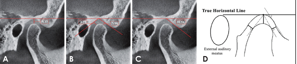
Fig. 1 A. Measurement of the superior joint space. B. The anterior and posterior joint spaces. C. The articular eminence inclination. D. A schematic view.
Cited by 1 articles
-
Reliability of cone-beam computed tomography for temporomandibular joint analysis
Hande Gorucu-Coskuner, Ezgi Atik, Hakan El
Korean J Orthod. 2019;49(2):81-88. doi: 10.4041/kjod.2019.49.2.81.
Reference
-
1. Abdel-Fattah RA. Optimum temporomandibular joint (TMJ) condylar position. Todays FDA. 1989; 1:1C–3C.2. Weffort SY, de Fantini SM. Condylar displacement between centric relation and maximum intercuspation in symptomatic and asymptomatic individuals. Angle Orthod. 2010; 80:835–842.
Article3. Bonilla-Aragon H, Tallents RH, Katzberg RW, Kyrkanides S, Moss ME. Condyle position as a predictor of temporomandibular joint internal derangement. J Prosthet Dent. 1999; 82:205–208.
Article4. Ilguy D, Ilguy M, Fisekcioglu E, Dolekoglu S, Ersan N. Articular eminence inclination, height, and condyle morphology on cone beam computed tomography. ScientificWorldJournal. 2014; 2014:761714.5. Arieta-Miranda JM, Silva-Valencia M, Flores-Mir C, Paredes-Sampen NA, Arriola-Guillen LE. Spatial analysis of condyle position according to sagittal skeletal relationship, assessed by cone beam computed tomography. Prog Orthod. 2013; 14:36.
Article6. Schiffman E, Ohrbach R, Truelove E, Look J, Anderson G, Goulet JP, et al. Diagnostic criteria for temporomandibular disorders (DC/TMD) for clinical and research applications: recommendations of the International RDC/TMD Consortium Network* and Orofacial Pain Special Interest Group. J Oral Facial Pain Headache. 2014; 28:6–27.7. Manfredini D, Guarda-Nardini L, Winocur E, Piccotti F, Ahlberg J, Lobbezoo F. Research diagnostic criteria for temporomandibular disorders: a systematic review of axis I epidemiologic findings. Oral Surg Oral Med Oral Pathol Oral Radiol Endod. 2011; 112:453–462.
Article8. Ren YF, Isberg A, Westesson PL. Condyle position in the temporomandibular joint. Comparison between asymptomatic volunteers with normal disk position and patients with disk displacement. Oral Surg Oral Med Oral Pathol Oral Radiol Endod. 1995; 80:101–107.9. Ronquillo HI, Guay J, Tallents RH, Katzberg RW, Murphy W. Tomographic analysis of mandibular condyle position as compared to arthrographic findings of the temporomandibular joint. J Craniomandib Disord. 1988; 2:59–64.10. Kurita H, Ohtsuka A, Kobayashi H, Kurashina K. A study of the relationship between the position of the condylar head and displacement of the temporomandibular joint disk. Dentomaxillofac Radiol. 2001; 30:162–165.
Article11. Rodrigues AF, Fraga MR, Vitral RW. Computed tomography evaluation of the temporomandibular joint in Class I malocclusion patients: condylar symmetry and condyle-fossa relationship. Am J Orthod Dentofacial Orthop. 2009; 136:192–198.
Article12. Vitral RW, Telles Cde S, Fraga MR, de Oliveira RS, Tanaka OM. Computed tomography evaluation of temporomandibular joint alterations in patients with class II division 1 subdivision malocclusions: condyle-fossa relationship. Am J Orthod Dentofacial Orthop. 2004; 126:48–52.
Article13. Ahmad M, Hollender L, Anderson Q, Kartha K, Ohrbach R, Truelove EL, et al. Research diagnostic criteria for temporomandibular disorders (RDC/TMD): development of image analysis criteria and examiner reliability for image analysis. Oral Surg Oral Med Oral Pathol Oral Radiol Endod. 2009; 107:844–860.
Article14. Tsiklakis K, Syriopoulos K, Stamatakis HC. Radiographic examination of the temporomandibular joint using cone beam computed tomography. Dentomaxillofac Radiol. 2004; 33:196–201.
Article15. McNeill C. Management of temporomandibular disorders: concepts and controversies. J Prosthet Dent. 1997; 77:510–522.
Article16. Martin D, Rozencweig S, Maté A, Valenzuela J. The importance of condyle position in the diagnosis, treatment and prevention of TMD. Orthod Fr. 2015; 86:125–149.17. Dalili Z, Khaki N, Kia SJ, Salamat F. Assessing joint space and condylar position in the people with normal function of temporomandibular joint with cone-beam computed tomography. . Dent Res J (Isfahan). 2012; 9:607–612.
Article18. Ikeda K, Kawamura A. Assessment of optimal condylar position with limited cone-beam computed tomography. Am J Orthod Dentofacial Orthop. 2009; 135:495–501.
Article19. Fernández Sanromán J, Gómez González JM, del Hoyo JA. Relationship between condylar position, dentofacial deformity and temporomandibular joint dysfunction: an MRI and CT prospective study. J Craniomaxillofac Surg. 1998; 26:35–42.
Article20. Christiansen EL, Thompson JR, Zimmerman G. Computed tomography of condylar and articular disk positions within the temporomandibular joint. Oral Surg Oral Med Oral Pathol. 1987; 64:757–767.
Article21. Al-koshab M, Nambiar P, John J. Assessment of condyle and glenoid fossa morphology using CBCT in South-East Asians. PLoS One. 2015; 10:e0121682.
Article22. Sulun T, Cemgil T, Duc JM, Rammelsberg P, Jager L, Gernet W. Morphology of the mandibular fossa and inclination of the articular eminence in patients with internal derangement and in symptom-free volunteers. Oral Surg Oral Med Oral Pathol Oral Radiol Endod. 2001; 92:98–107.23. Kurita H, Ohtsuka A, Kobayashi H, Kurashina K. Flattening of the articular eminence correlates with progressive internal derangement of the temporomandibular joint. Dentomaxillofac Radiol. 2000; 29:277–279.
Article24. Incesu L, Taşkaya-Yılmaz N, Öğütcen-Toller M, Uzun E. Relationship of condylar position to disc position and morphology. Eur J Radiol. 2004; 51:269–273.
Article25. Gateno J, Anderson PB, Xia JJ, Horng JC, Teichgraeber JF, Liebschner MA. A comparative assessment of mandibular condylar position in patients with anterior disc displacement of the temporomandibular joint. J Oral Maxillofac Surg. 2004; 62:39–43.
Article26. Kinniburgh RD, Major PW, Nebbe B, West K, Glover KE. Osseous morphology and spatial relationships of the temporomandibular joint: comparisons of normal and anterior disc positions. Angle Orthod. 2000; 70:70–78.
- Full Text Links
-
- Actions
-
Cited
- CITED
-
- Close
- Share
-
- Similar articles
-
- Osteoarthritic changes and condylar positioning of the temporomandibular joint in Korean children and adolescents
- Cone-beam computed tomographic evaluation of the condylar remodeling occurring after mandibular set-back by bilateral sagittal split ramus osteotomy and rigid fixation
- Three-dimensional cone-beam computed tomography based comparison of condylar position and morphology according to the vertical skeletal pattern
- Mandibular condyle position in cone beam computed tomography
- Three-dimensional assessment of the temporomandibular joint and mandibular dimensions after early correction of the maxillary arch form in patients with Class II division 1 or division 2 malocclusion
