Korean J Endocr Surg.
2015 Dec;15(4):99-102. 10.16956/kaes.2015.15.4.99.
A Novel Germline Mutation of MEN1 Gene in a Young-aged Multiple Insulinoma with Hyperparathyroidism
- Affiliations
-
- 1Department of Surgery, Dankook University College of Medicine, Cheonan, Korea. psurgeon@naver.com
- KMID: 2308577
- DOI: http://doi.org/10.16956/kaes.2015.15.4.99
Abstract
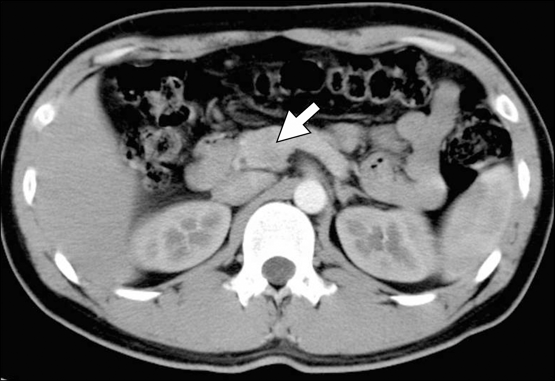
- Multiple endocrine neoplasia type 1 is an autosomal dominant disease caused by the MEN1 germline mutation. A 25-year-old male was admitted for loss of consciousness. Initial laboratory data showed hypoglycemia and hypercalcemia. The image study showed two insulinoma in the pancreas head and body. MIBI scan was positive in the left lower parathyroid gland. After diagnosis of insulinoma and hyperparathyroidism, MEN1 was suspected, but there was no family history of endocrine disease. Enucleation of the insulinoma in the pancreatic head and body was performed. After the operation, the blood sugar level was normalized and no hypoglycemic symptoms were observed. Testing of germline mutations of the MEN1 gene was performed by direct DNA sequence analysis after obtaining informed consent. In the genetic study, a frameshift mutation was found in exon 2 which deleted 16 nucleic acids (c.326_341del16) and resulted in a truncation at codon 113. This mutation was not reported previously. We found a novel and de novo mutation of the MEN1 gene. Genetic study is necessary in case of young-age, multiple endocrine tumors.
MeSH Terms
-
Adult
Blood Glucose
Codon
Diagnosis
Endocrine System Diseases
Exons
Frameshift Mutation
Germ-Line Mutation*
Head
Humans
Hypercalcemia
Hyperparathyroidism*
Hypoglycemia
Informed Consent
Insulinoma*
Male
Multiple Endocrine Neoplasia Type 1*
Nucleic Acids
Pancreas
Parathyroid Glands
Sequence Analysis, DNA
Unconsciousness
Blood Glucose
Codon
Nucleic Acids
Figure
Reference
-
References
1. Romei C, Pardi E, Cetani F, Elisei R. Genetic and clinical features of multiple endocrine neoplasia types 1 and 2. J Oncol. 2012; 2012:705036.
Article2. Bergman L, Teh B, Cardinal J, Palmer J, Walters M, Shepherd J, et al. Identification of MEN1 gene mutations in families with MEN 1 and related disorders. Br J Cancer. 2000; 83:1009–14.
Article3. Cohen MS, Picus D, Lairmore TC, Strasberg SM, Doherty GM, Norton JA. Prospective study of provocative angiograms to localize functional islet cell tumors of the pancreas. Surgery. 1997; 122:1091–100.
Article4. Doherty GM, Doppman JL, Shawker TH, Miller DL, Eastman RC, Gorden P, et al. Results of a prospective strategy to diagnose, localize, and resect insulinomas. Surgery. 1991; 110:989–96. discussion 996–7.5. Bilezikian JP, Khan AA. Potts JT Jr; Third International Workshop on the Management of Asymptomatic Primary Hyperthyroidism. Guidelines for the management of asymptomatic primary hyperparathyroidism: summary statement from the third international workshop. J Clin Endocrinol Metab. 2009; 94:335–9.
Article6. Lemos MC, Thakker RV. Multiple endocrine neoplasia type 1 (MEN1): analysis of 1336 mutations reported in the first decade following identification of the gene. Hum Mutat. 2008; 29:22–32.
Article7. Lee SC, Min JW, Kim YM, Chang MC. Characteristics of the germline MEN1 mutations in Korea: a literature review. Korean J Endocr Surg. 2014; 14:7–11.
Article
- Full Text Links
- Actions
-
Cited
- CITED
-
- Close
- Share
- Similar articles
-
- A Case of Familial Multiple Endocrine Neoplasia Type 1 with a Novel Mutation in the MEN1 Gene
- A Case of Familial Isolated Primary Hyperparathyroidism with a Novel Gene Mutation
- A Case of Familial Multiple Endocrine Neoplasia with MEN1 Gene Mutation
- Characteristics of the Germline MEN1 Mutations in Korea: A Literature Review
- Clinical Manifestation of Multiple Endocrine Neoplasia Type I


